

Realistic simulation of the 3-D growth of brain tumors in MR images coupling diffusion with biomechanical deformation
- PMID: 16229419
- PMCID: PMC2496876
- DOI: 10.1109/TMI.2005.857217
Realistic simulation of the 3-D growth of brain tumors in MR images coupling diffusion with biomechanical deformation
Abstract
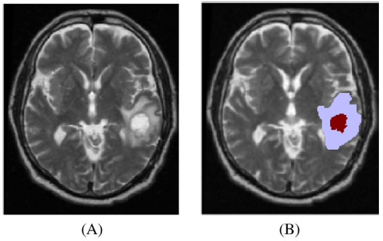
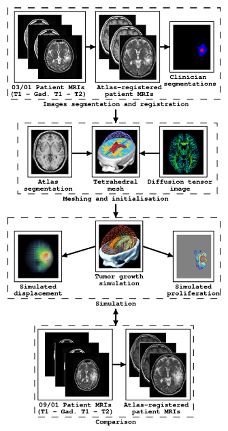
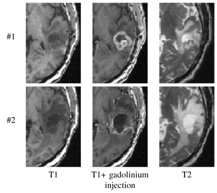
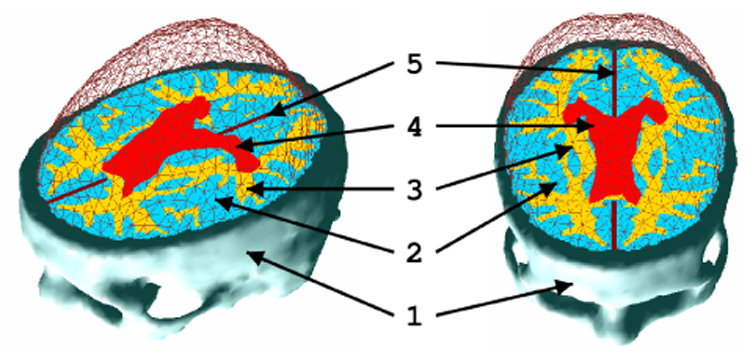
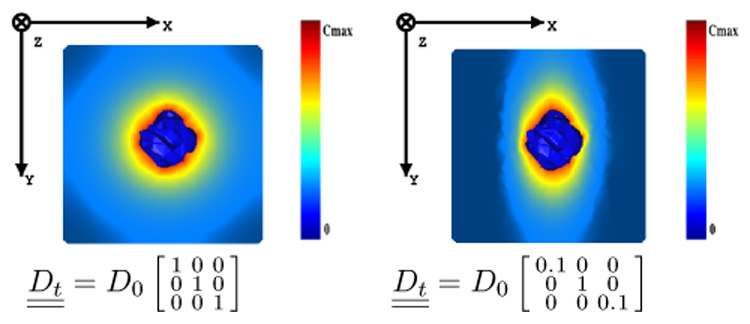
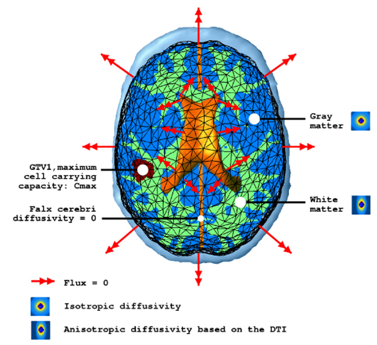
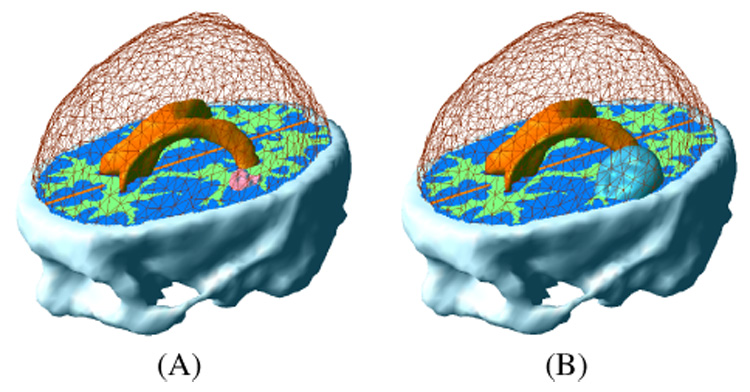
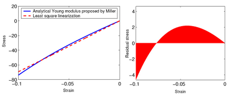
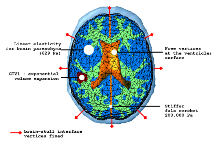
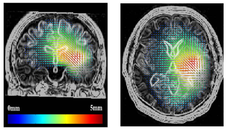
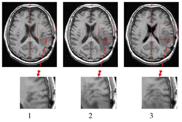
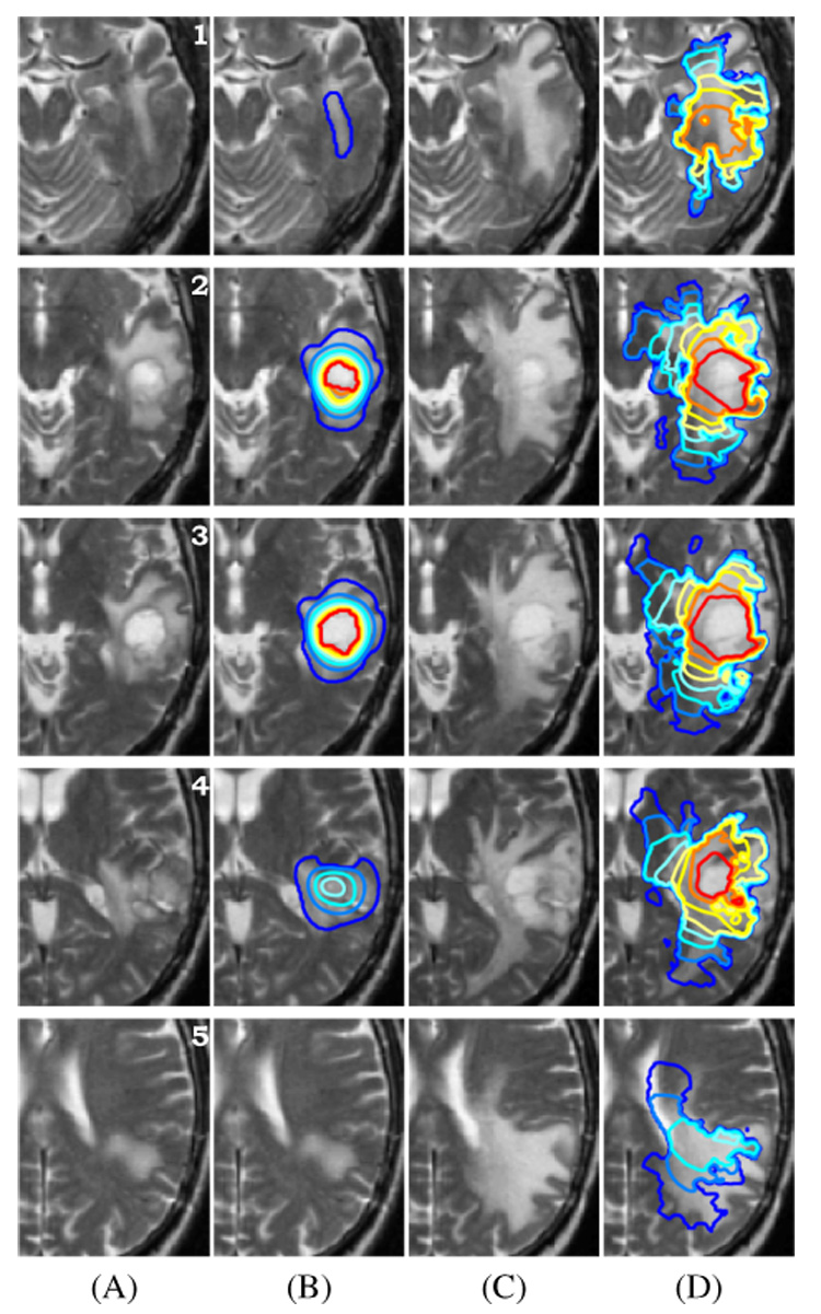
We propose a new model to simulate the three-dimensional (3-D) growth of glioblastomas multiforma (GBMs), the most aggressive glial tumors. The GBM speed of growth depends on the invaded tissue: faster in white than in gray matter, it is stopped by the dura or the ventricles. These different structures are introduced into the model using an atlas matching technique. The atlas includes both the segmentations of anatomical structures and diffusion information in white matter fibers. We use the finite element method (FEM) to simulate the invasion of the GBM in the brain parenchyma and its mechanical interaction with the invaded structures (mass effect). Depending on the considered tissue, the former effect is modeled with a reaction-diffusion or a Gompertz equation, while the latter is based on a linear elastic brain constitutive equation. In addition, we propose a new coupling equation taking into account the mechanical influence of the tumor cells on the invaded tissues. The tumor growth simulation is assessed by comparing the in-silico GBM growth with the real growth observed on two magnetic resonance images (MRIs) of a patient acquired with 6 mo difference. Results show the feasibility of this new conceptual approach and justifies its further evaluation.
Figures
References
-
- Kantor G, Loiseau H, Vital A, Mazeron JJ. Gross tumor volume (gtv) and clinical target volume (ctv) in adult gliomas. Cancer Radiother. 2001 October;vol. 5(no. 5):571–580. - PubMed
-
- Retsky MW, Swartzendruber DE, Wardwell RH, Bame PD. Is gompertzian or exponential kinetics a valid description of individual human cancer growth? Med Hypotheses. 1990 October;vol. 33(no. 2):95–106. - PubMed
-
- Lazareff JA, Suwinski R, De Rosa R, De RR, Olmstead CE. Tumor volume and growth kinetics in hypothalamic-chiasmatic pediatric low grade gliomas. Pediatr Neurosurg. 1999 June;vol. 30(no. 6):312–319. - PubMed
-
- Bajzer Z. Gompertzian growth as a self-similar and allometric process. Growth Dev Aging. 1999 Spring-Summer;vol. 63(no. 1–2):3–11. - PubMed
Publication types
MeSH terms
Grants and funding
LinkOut - more resources
Full Text Sources
Other Literature Sources
Medical
