. 2005 Sep;7(9):799-808.
doi: 10.1593/neo.05274.

Epigenetic regulation of the cell type-specific gene 14-3-3sigma

Affiliations

Epigenetic regulation of the cell type-specific gene 14-3-3sigma

Marc M Oshiro et al. Neoplasia. 2005 Sep.

Abstract

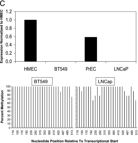
Epigenetic control participates in processes crucial in mammalian development, such as X-chromosome inactivation, gene imprinting, and cell type-specific gene expression. We provide evidence that the p53-inducible gene 14-3-3sigma is a new example of a gene important to human cancer, where epigenetic mechanisms participate in the control of normal cell type-specific expression, as well as aberrant gene silencing in cancer cells. Like a previously identified cell type-specific gene maspin, 14-3-3sigma is a p53-inducible gene; however, it participates in G2/M arrest in response to DNA-damaging agents. 14-3-3Sigma expression is restricted to certain epithelial cell types, including breast and prostate, whereas expression is absent in nonepithelial tissues such as fibroblasts and lymphocytes. In this report, we show that in normal cells expressing 14-3-3sigma, the 14-3-3sigma CpG island is unmethylated; associated with acetylated histones, unmethylated histone H3 lysine 9; and an accessible chromatin structure. By contrast, normal cells that do not express 14-3-3sigma have a methylated 14-3-3sigma CpG island with hypoacetylated histones, methylated histone H3 lysine 9, and an inaccessible chromatin structure. These findings extend the spectrum of cell type-specific genes controlled, partly, by normal epigenetic mechanisms, and suggest that this subset of genes may represent important targets of epigenetic dysregulation in human cancer.


Figures

Figure 1
Figure 1
14-3-3σ Expression is restricted to a subset of normal human cell types. 14-3-3σ Expression was assessed by real-time quantitative RT-PCR; GAPDH was used to normalize the expression data.
Figure 2
Figure 2
The 14-3-3σ CpG island shows differential cytosine methylation between 14-3-3σ-positive and 14-3-3σ-negative normal human tissue. (A) Diagram of the 14-3-3σ gene and CpG island region analyzed. CpG dinucleotide, MspI restriction sites, and primers utilized are demarcated on the diagram. (B) Summary of 5-methylcytosine levels in the 14-3-3σ CpG island. Ten cloned PCR products were sequenced to determine the percent methylation of the 27 CpG sites. (C) Tumor cells are able to recapitulate methylation-induced gene silencing analogous to normal 14-3-3σ-negative cell types. RNA was isolated and subjected to real-time PCR analysis of 14-3-3σ, and bisulfite-modified DNA was sequenced for analysis of methylation.
Figure 2
Figure 2
The 14-3-3σ CpG island shows differential cytosine methylation between 14-3-3σ-positive and 14-3-3σ-negative normal human tissue. (A) Diagram of the 14-3-3σ gene and CpG island region analyzed. CpG dinucleotide, MspI restriction sites, and primers utilized are demarcated on the diagram. (B) Summary of 5-methylcytosine levels in the 14-3-3σ CpG island. Ten cloned PCR products were sequenced to determine the percent methylation of the 27 CpG sites. (C) Tumor cells are able to recapitulate methylation-induced gene silencing analogous to normal 14-3-3σ-negative cell types. RNA was isolated and subjected to real-time PCR analysis of 14-3-3σ, and bisulfite-modified DNA was sequenced for analysis of methylation.
Figure 3
Figure 3
The histone acetylation state of the 14-3-3σ CpG island is associated with cytosine methylation and expression status in normal human tissue. Acetylation of histones H3 and H4 in the 14-3-3σ CpG island and GAPDH promoter was analyzed using a quantitative ChIP assay coupled to real-time PCR. Chromatin was immunoprecipitated from the normal cell types with either an acetylation-specific H3 or H4 antibody, or no antibody. Real-time PCR was performed by utilizing primer/probe sets specific for the 14-3-3σ CpG island and to the promoter region of GAPDH. HFFs, human foreskin fibroblasts; HMECs, human mammary epithelial cells; PrECs, prostate epithelial cells; PBLs, peripheral blood lymphocytes.
Figure 4
Figure 4
The histone H3 dimethyl-lysine 9 methylation state of the 14-3-3σ CpG island is associated with cytosine methylation, histone acetylation, and expression status in normal human tissues. Methylation of histone H3 lysine 9 within the 14-3-3σ CpG island and GAPDH promoter was analyzed using a quantitative ChIP assay. Chromatin was immunoprecipitated from the normal cell types with a dimethyl histone H3 lysine 9 antibody or no antibody. Real-time PCR was performed by utilizing primers specific for the 14-3-3σ CpG island and to the promoter region of GAPDH.
Figure 5
Figure 5
Chromatin structure of the 14-3-3σ CpG island in positive and negative cell types correlate with unmethylated, 14-3-3σ-expressing and methylated, 14-3-3σ-nonexpressing tissue types, respectively. Intact nuclei were digested in vivo with MspI restriction endonuclease. Following in vivo digestion, gDNA was isolated. Real-time SYBR Green PCR was performed using primers that flank five MspI sites. Inaccessible sites are shown by a positive display of a PCR product, whereas accessible sites that are digested by MspI yield fewer products following amplification. As a positive control for PCR amplification, an equal amount of uncut gDNA was assayed; as a negative control, an equal amount of gDNA digested with MspI was assayed.
Figure 6
Figure 6
DNA damage induction of 14-3-3σ is not induced in normal epigenetically silenced 14-3-3σ cell types. (A) HFFs were treated with doxorubicin for 18 hours prior to collection of protein whole cell lysates. Western blot analysis for p53 was conducted. (B) HFFs were treated with doxorubicin for 18 hours prior to collection of total RNA. Real-time quantitative RT-PCR was conducted to analyze the expression of the p53-inducible genes 14-3-3σ, p21, and MDM2. PrECs served as an example of a normal 14-3-3σ-positive cell type.

Similar articles

Cited by

References

    1. Li E, Bestor TH, Jaenisch R. Targeted mutation of the DNA methyltransferase gene results in embryonic lethality. Cell. 1992;69:915–926. - PubMed
    1. Gardiner-Garden M, Frommer M. CpG islands in vertebrate genomes. J Mol Biol. 1987;196:261–282. - PubMed
    1. Takai D, Jones PA. The CpG island searcher: a new WWW resource. In Silico Biol. 2003;3:235–240. - PubMed
    1. Ehrlich M. DNA methylation in cancer: too much, but also too little. Oncogene. 2002;21:5400–5413. - PubMed
    1. Feinberg AP, Tycko B. The history of cancer epigenetics. Nat Rev Cancer. 2004;4:143–153. - PubMed

Publication types

MeSH terms

LinkOut - more resources