

I.V. infusion of brain-derived neurotrophic factor gene-modified human mesenchymal stem cells protects against injury in a cerebral ischemia model in adult rat
- PMID: 16229956
- PMCID: PMC2605391
- DOI: 10.1016/j.neuroscience.2005.06.062
I.V. infusion of brain-derived neurotrophic factor gene-modified human mesenchymal stem cells protects against injury in a cerebral ischemia model in adult rat
Abstract
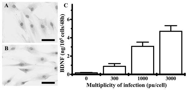
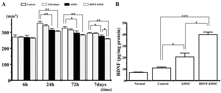
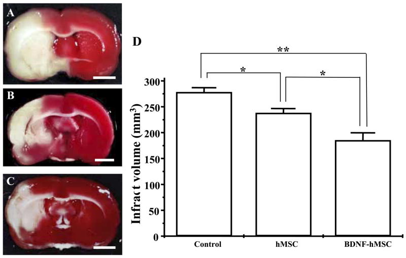
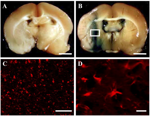
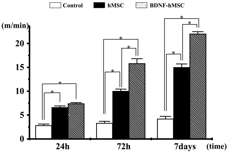
I.V. delivery of mesenchymal stem cells prepared from adult bone marrow reduces infarction size and ameliorates functional deficits in rat cerebral ischemia models. Administration of the brain-derived neurotrophic factor to the infarction site has also been demonstrated to be neuroprotective. To test the hypothesis that brain-derived neurotrophic factor contributes to the therapeutic benefits of mesenchymal stem cell delivery, we compared the efficacy of systemic delivery of human mesenchymal stem cells and human mesenchymal stem cells transfected with a fiber-mutant F/RGD adenovirus vector with a brain-derived neurotrophic factor gene (brain-derived neurotrophic factor-human mesenchymal stem cells). A permanent middle cerebral artery occlusion was induced by intraluminal vascular occlusion with a microfilament. Human mesenchymal stem cells and brain-derived neurotrophic factor-human mesenchymal stem cells were i.v. injected into the rats 6 h after middle cerebral artery occlusion. Lesion size was assessed at 6 h, 1, 3 and 7 days using MR imaging, and histological methods. Functional outcome was assessed using the treadmill stress test. Both human mesenchymal stem cells and brain-derived neurotrophic factor-human mesenchymal stem cells reduced lesion volume and elicited functional improvement compared with the control sham group, but the effect was greater in the brain-derived neurotrophic factor-human mesenchymal stem cell group. ELISA analysis of the infarcted hemisphere revealed an increase in brain-derived neurotrophic factor in the human mesenchymal stem cell groups, but a greater increase in the brain-derived neurotrophic factor-human mesenchymal stem cell group. These data support the hypothesis that brain-derived neurotrophic factor contributes to neuroprotection in cerebral ischemia and cellular delivery of brain-derived neurotrophic factor can be achieved by i.v. delivery of human mesenchymal stem cells.
Figures

References
-
- Alvarez-Dolado M, Pardal R, Garcia-Verdugo JM, Fike JR, Lee HO, Pfeffer K, Lois C, Morrison SJ, Alvarez-Buylla A. Fusion of bone-marrow-derived cells with Purkinje neurons, cardiomyocytes and hepatocytes. Nature. 2003;425:968–973. - PubMed
-
- Bederson JB, Pitts LH, Germano SM, Nishimura MC, Davis RL, Bartkowski HM. Evaluation of 2,3,5-triphenyltetrazolium chloride as a stain for detection and quantification of experimental cerebral infarction in rats. Stroke. 1986;17:1304–1308. - PubMed
-
- Brazelton TR, Rossi FM, Keshet GI, Blau HM. From marrow to brain: expression of neuronal phenotypes in adult mice. Science. 2000;290:1775–1779. - PubMed
-
- Castro RF, Jackson KA, Goodell MA, Robertson CS, Liu H, Shine HD. Failure of bone marrow cells to transdifferentiate into neural cells in vivo. Science. 2002;23:297. - PubMed
Publication types
MeSH terms
Substances
Grants and funding
LinkOut - more resources
Full Text Sources
Other Literature Sources
