Comparative Study
. 2005 Oct 17;202(8):1043-9.
doi: 10.1084/jem.20050977.

Innate immunity against Francisella tularensis is dependent on the ASC/caspase-1 axis

Affiliations
Comparative Study

Innate immunity against Francisella tularensis is dependent on the ASC/caspase-1 axis

Sanjeev Mariathasan et al. J Exp Med. .

Abstract

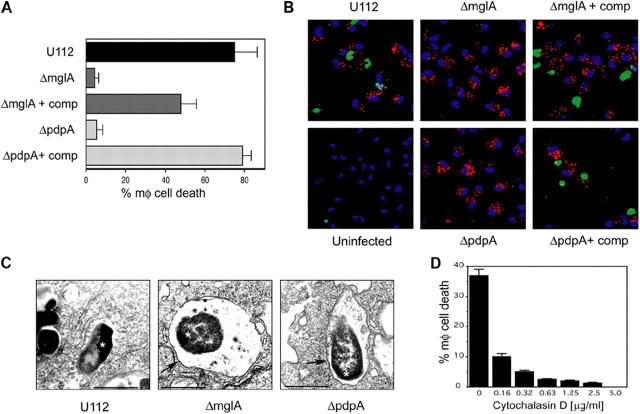
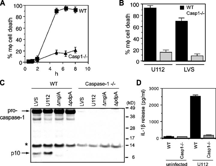
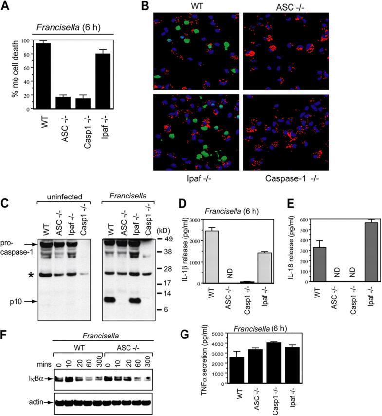
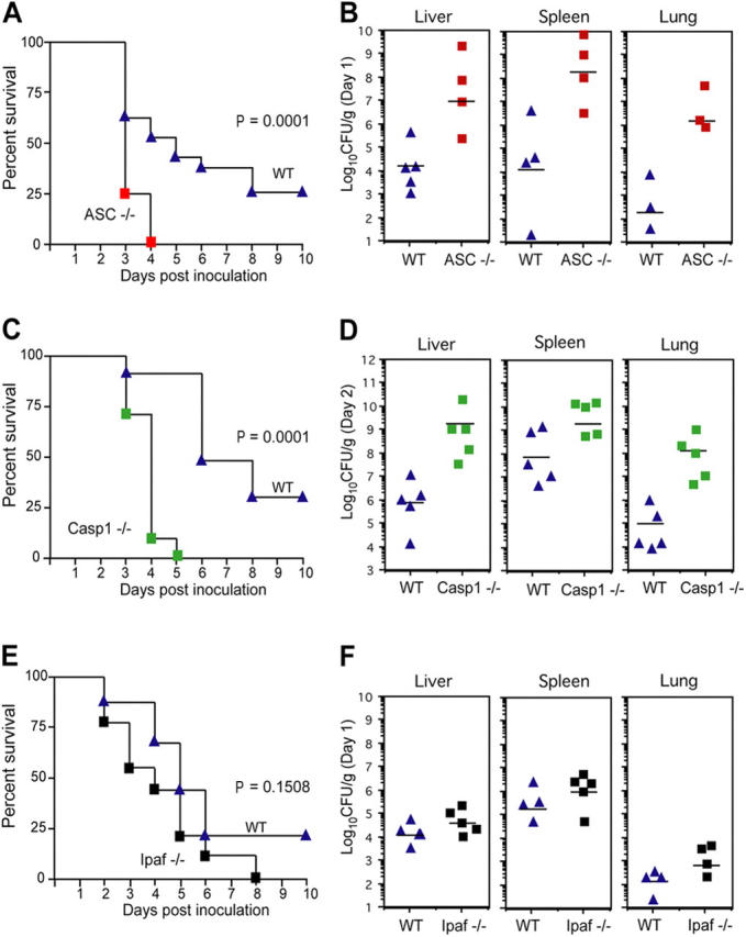
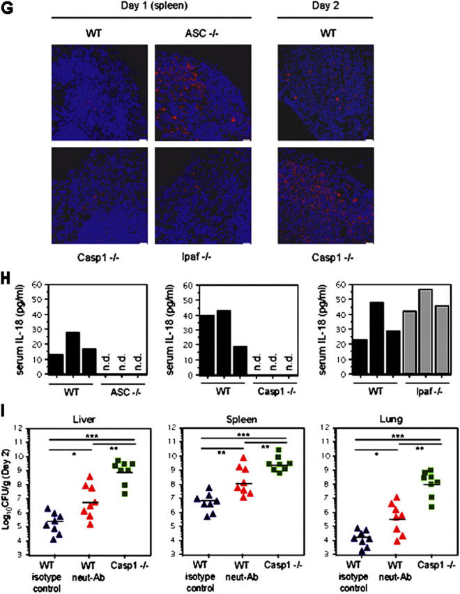
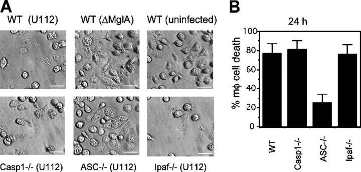
Francisella tularensis is a highly infectious gram-negative coccobacillus that causes the zoonosis tularemia. This bacterial pathogen causes a plague-like disease in humans after exposure to as few as 10 cells. Many of the mechanisms by which the innate immune system fights Francisella are unknown. Here we show that wild-type Francisella, which reach the cytosol, but not Francisella mutants that remain localized to the vacuole, induced a host defense response in macrophages, which is dependent on caspase-1 and the death-fold containing adaptor protein ASC. Caspase-1 and ASC signaling resulted in host cell death and the release of the proinflammatory cytokines interleukin (IL)-1beta and IL-18. F. tularensis-infected caspase-1- and ASC-deficient mice showed markedly increased bacterial burdens and mortality as compared with wild-type mice, demonstrating a key role for caspase-1 and ASC in innate defense against infection by this pathogen.


Figures

Figure 1.
Figure 1.
Francisella-induced macrophage death requires bacterial localization to the cytosol and the mglA and pdpA genes. Macrophages were infected with wild-type Francisella U112 (WT; multiplicity of infection [moi] 30), ΔmglA (moi 200), ΔmglA + mglA (moi 30), ΔpdpA (moi 200), ΔpdpA + pdpA (moi 30), and cell death was measured by (A) LDH release during the 8-h infection (% mφ cell death) or (B) TUNEL. Macrophages were fixed 4 h after infection and stained with TUNEL (green), chicken anti-Francisella primary antibody, anti–chicken-alexa594 secondary antibody (red), and the TOTO-3 DNA stain (blue). (C) Macrophages were infected with WT, ΔmglA, or ΔpdpA for 6 h, fixed, and processed for transmission electron microscopy. Asterisks (*) denote intracellular bacteria and arrows point to phagosomal membranes. WT bacteria are not surrounded by a phagosomal membrane. Bar, 0.7 μm. (D) Macrophages were pretreated for 1 h with the indicated concentrations of cytochalasin D, washed, and infected with WT (moi 30) for 5 h followed by measurement of LDH release. Samples were performed in triplicate. Experiments were performed at least three times. Means and standard deviations from a representative experiment are shown.
Figure 2.
Figure 2.
Casp-1 is essential for early F. tularensis–induced macrophage death. WT and casp-1 −/− macrophages were infected (moi 30) with F. tularensis U112 (A and B) or LVS (B) for 8 h. Cell death was measured by LDH release into the culture supernatant (% mφ cell death). (C) Macrophages were infected with LVS (moi 30), U112 (moi 30), ΔmglA (moi 200) or ΔpdpA (moi 200) and cell lysates were collected at 4 h and immunoblotted with antibodies against the p10 subunit of casp-1. The asterisk (*) denotes a nonspecific cross-reactive band. (D) Macrophages were infected with U112 (moi 30) and IL-1β release into the supernatant at 6 h was measured by ELISA. Samples were performed in triplicate. Experiments were performed at least three times. Means and standard deviations from a representative experiment are shown.
Figure 3.
Figure 3.
ASC is essential for Francisella-induced casp-1 activation and macrophage death. Macrophages from WT, Asc −/−, Ipaf −/−, or casp-1 −/− mice were infected with WT Francisella U112 (moi 30) for 6 h. (A) Cell death was monitored by LDH release (% mφ cell death). (B) 6 h p.i., macrophages were fixed and stained with TUNEL (green), chicken anti-Francisella primary antibody, anti–chicken-alexa594 secondary antibody (red), and TOTO-3 DNA stain (blue). (C) Cell lysates were immunoblotted with antibodies against the p10 subunit of casp-1. The asterisk (*) denotes a nonspecific cross-reactive band. (D and E) IL-1β and IL-18 release into the supernatant was measured by ELISA. (F) Analysis of IκB degradation in Francisella-infected macrophages. Macrophages were infected with WT U112 for 6 h. Cell lysates were analyzed by Western blot using anti-phosphoIκB antibody (S32; top) or antiactin antibody (bottom). (G) TNFα release into the supernatant was measured by ELISA. Western blot analyses were performed at least three times and representative blots are shown. ELISAs were performed in triplicate, means and standard deviations are shown.
Figure 4.
Figure 4.
ASC and casp-1 are essential for host defense against F. tularensis in vivo. (A, C, and E) Mice were injected subcutaneously with WT Francisella U112 (1.5 × 10⁵ CFU) and their survival was monitored (Asc / n = 22, Asc + / + n = 23; casp-1 / n = 10, casp-1 + / + n = 10; Ipaf / n = 9, Ipaf + / + n = 9) or (B, D, and F) tissues were recovered 1 or 2 d p.i., homogenized, and dilutions plated on bacterial media for enumeration of CFU. Bacterial counts on day 1 from the liver, spleen, and lung of Asc / mice were significantly higher compared with WT mice (P = 0.0317, 0.0471, and 0.0159, respectively). Day 2 counts from casp-1 / mice were also significantly higher compared with WT mice (liver; P = 0.0079; spleen, P = 0.0317; lung, P = 0.0079). (G) Spleen sections were labeled with anti-Francisella antibody (red) and TOTO-3 (blue) to stain all nuclei. The image is a projection of an 8-μm z-stack collected through the 60× objective on a confocal microscope. Bar, 20 μm. (H) Levels of IL-18 in the serum of mice 1 d p.i. were determined by ELISA. (I) WT mice were intraperitoneally injected with neutralizing IL-18 and IL-1β antibodies (n = 8) or control antibodies (n = 8). 30 min later, antibody-treated mice, as well as untreated casp-1 / mice (n = 8), were subcutaneously injected with 105 CFU of strain U112. 2 d p.i., tissues were collected, homogenized, and dilutions were plated on bacterial media to count CFU. *, P < 0.05; **, P < 0.005; ***, P < 0.0005. All experiments were performed at least three times.
Figure 4.
Figure 4.
ASC and casp-1 are essential for host defense against F. tularensis in vivo. (A, C, and E) Mice were injected subcutaneously with WT Francisella U112 (1.5 × 10⁵ CFU) and their survival was monitored (Asc / n = 22, Asc + / + n = 23; casp-1 / n = 10, casp-1 + / + n = 10; Ipaf / n = 9, Ipaf + / + n = 9) or (B, D, and F) tissues were recovered 1 or 2 d p.i., homogenized, and dilutions plated on bacterial media for enumeration of CFU. Bacterial counts on day 1 from the liver, spleen, and lung of Asc / mice were significantly higher compared with WT mice (P = 0.0317, 0.0471, and 0.0159, respectively). Day 2 counts from casp-1 / mice were also significantly higher compared with WT mice (liver; P = 0.0079; spleen, P = 0.0317; lung, P = 0.0079). (G) Spleen sections were labeled with anti-Francisella antibody (red) and TOTO-3 (blue) to stain all nuclei. The image is a projection of an 8-μm z-stack collected through the 60× objective on a confocal microscope. Bar, 20 μm. (H) Levels of IL-18 in the serum of mice 1 d p.i. were determined by ELISA. (I) WT mice were intraperitoneally injected with neutralizing IL-18 and IL-1β antibodies (n = 8) or control antibodies (n = 8). 30 min later, antibody-treated mice, as well as untreated casp-1 / mice (n = 8), were subcutaneously injected with 105 CFU of strain U112. 2 d p.i., tissues were collected, homogenized, and dilutions were plated on bacterial media to count CFU. *, P < 0.05; **, P < 0.005; ***, P < 0.0005. All experiments were performed at least three times.
Figure 5.
Figure 5.
ASC is required for two stages of macrophage death in response to Francisella. Macrophages from WT, Asc −/−, casp-1 −/−, or Ipaf −/− mice were infected with F. tularensis U112 (moi 30) for 24 h and (A) imaged by differential interference contrast microscopy or (B) assayed for macrophage death by LDH release (% mφ cell death). Samples were performed in triplicate. Experiments were performed at least three times. Means and standard deviations from a representative experiment are shown.

References

    1. Sjostedt, A. 2003. Virulence determinants and protective antigens of Francisella tularensis. Curr. Opin. Microbiol. 6:66–71. - PubMed
    1. Elkins, K.L., S.C. Cowley, and C.M. Bosio. 2003. Innate and adaptive immune responses to an intracellular bacterium, Francisella tularensis live vaccine strain. Microbes Infect. 5:135–142. - PubMed
    1. Lindgren, H., S. Stenmark, W. Chen, A. Tarnvik, and A. Sjostedt. 2004. Distinct roles of reactive nitrogen and oxygen species to control infection with the facultative intracellular bacterium Francisella tularensis. Infect. Immun. 72:7172–7182. - PMC - PubMed
    1. Nano, F.E., N. Zhang, S.C. Cowley, K.E. Klose, K.K. Cheung, M.J. Roberts, J.S. Ludu, G.W. Letendre, A.I. Meierovics, G. Stephens, and K.L. Elkins. 2004. A Francisella tularensis pathogenicity island required for intramacrophage growth. J. Bacteriol. 186:6430–6436. - PMC - PubMed
    1. Lauriano, C.M., J.R. Barker, S.S. Yoon, F.E. Nano, B.P. Arulanandam, D.J. Hassett, and K.E. Klose. 2004. MglA regulates transcription of virulence factors necessary for Francisella tularensis intraamoebae and intramacrophage survival. Proc. Natl. Acad. Sci. USA. 101:4246–4249. - PMC - PubMed

Publication types

MeSH terms