. 2005 Nov 1;19(21):2571-82.
doi: 10.1101/gad.1348905. Epub 2005 Oct 17.

A histone code in meiosis: the histone kinase, NHK-1, is required for proper chromosomal architecture in Drosophila oocytes

Affiliations

A histone code in meiosis: the histone kinase, NHK-1, is required for proper chromosomal architecture in Drosophila oocytes

Irena Ivanovska et al. Genes Dev. .

Abstract

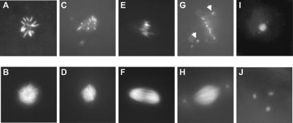
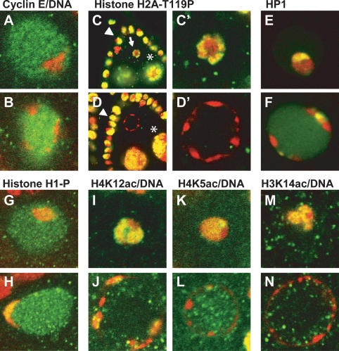
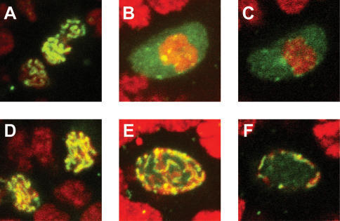
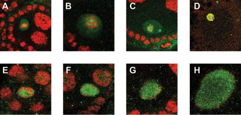
To promote faithful propagation of the genetic material during sexual reproduction, meiotic chromosomes undergo specialized morphological changes that ensure accurate segregation of homologous chromosomes. The molecular mechanisms that establish the meiotic chromosomal structures are largely unknown. We describe a mutation in a recently identified Histone H2A kinase, nhk-1, in Drosophila that leads to female sterility due to defects in the formation of the meiotic chromosomal structures. The metaphase I arrest and the karyosome, a critical prophase I chromosomal structure, require nucleosomal histone kinase-1 (NHK-1) function. The defects are a result of failure to disassemble the synaptonemal complex and to load condensin onto the mutant chromosomes. Embryos laid by nhk-1-/- mutant females arrest with aberrant polar bodies and mitotic spindles, revealing that mitosis is affected as well. We analyzed the role of Histone H2A phosphorylation with respect to the histone code hypothesis and found that it is required for acetylation of Histone H3 and Histone H4 in meiosis. These studies reveal a critical role for histone modifications in chromosome dynamics in meiosis and mitosis.


Figures

Figure 1.
Figure 1.
The nhk-1Z3-0437 mutation leads to aberrant chromosomal arrangements in the polar body, the mitotic spindle, and the metaphase I spindle. (A) Normal polar body, or rosette, in a control embryo stained with DAPI. (B) Normal microtubule arrangement in a control embryo. (C) The chromosomes in some embryos laid by nhk-1Z3-0437 mutant females formed aberrant rosettes. (D) Microtubules surrounding the abnormal rosette in C had a normal appearance. (E) The chromosomes in other embryos laid by nhk-1Z3-0437 mutant females appeared aligned on a spindle plate. (F) Microtubules surrounding the chromosomes in E formed a spindle that lacked asters. (G) Embryos laid by nhk-1Z3-0437 mutant females had one or two additional chromosomal arrangements that represented the female and male pronuclei with the majority of chromosomes at a mitotic spindle plate. Some chromosomes in these spindles failed to align at the plate and were found near the spindle poles (arrows). (H) The microtubules surrounding the aberrant mitotic chromosomes appeared normal and had asters, suggesting that the embryos were fertilized. (I) Normal metaphase I chromosome configuration in control stage 14 oocytes.(J) The chromosomes in nhk-1Z3-0437 mutant stage 14 oocytes failed to align at a metaphase I spindle and were found in three distinct foci.
Figure 2.
Figure 2.
nhk-1Z3-0437 is a mutation in a histone kinase. (A, top) A schematic of the NHK-1 protein structure. The black bar represents the kinase domain and the dark gray bar represents the basic-acidic-basic (BAB) domain conserved among the closest homologs of the NHK-1 (Aihara et al. 2004). The approximate position of the Z3-0437 Pro 117-to-leucine mutation is indicated. (Bottom) The protein sequence of a portion of the kinase domain of NHK-1 and its homologs, showing the Pro 117 (bold) mutated in the Z3-0473 mutant and just upstream of the catalytic loop important for kinase activity. (B) nhk-1 transcript is present in wild-type Drosophila ovaries—signal from antisense-strand probe. (C) The sense-strand probe shows the level of background signal. (D, top) Ovarian extracts were probed with anti-NHK-1 antibodies. The left lane is an extract from nhk-1Z3-0437/+ heterozygous control ovaries and shows that NHK-1 protein is present in the ovaries. The right lane is from nhk-1Z3-0437/Df mutant ovaries and shows a slight reduction in the NHK-1 protein levels due to the mutation. (Bottom) The same blot was stripped and reprobed with anti-α-tubulin antibodies as a control for equal loading of the extracts.
Figure 3.
Figure 3.
NHK-1 and the histone code. (A) The karyosome (stained with propidium iodide, red) is a chromosomal structure that forms within the prophase I nucleus (stained with Cyclin E, green) in control nhk-1Z3-0437/+ oocytes. (B) nhk-1Z3-0437/Z3-0437 mutant oocytes do not form a karyosome. Instead, the DNA (red) is found at the periphery of the oocyte nucleus (green). (C) Histone H2A-T119ph (green) is present in nurse cells (), follicle cells (arrowhead), and the karyosomes (arrow) in control nhk-1Z3-0437/+ heterozygous ovaries, and it colocalizes with the DNA (red). Yellow denotes overlap between the signals. (C′) The control karyosome at a higher magnification showing the colocalization of Histone H2AT119ph with the DNA. (D) Histone H2A-T119ph persists in nurse cells () and follicle cells (arrowhead) in the mutant nhk-1Z3-0437/Z3-0437 ovaries. However, the DNA in the nhk-1Z3-0437/Z3-0437 mutant oocyte nucleus lacks Histone H2AT119ph. (D′) A higher magnification showing the absence of H2AT119ph in the mutant oocyte nucleus. (E) Heterochromatin protein 1 (HP1, green) localizes to the karyosome (red) in control nhk-1Z3-0437/+ oocytes. (F) HP1 colocalizes with the DNA in the nhk-1Z3-0437/Z3-0437 mutant oocytes. (G) Histone H1 is phosphorylated in the control nhk-1Z3-0437/+ karyosomes (DNA, red). (H) Phosphorylated Histone H1 colocalizes with the DNA (red) in the nhk-1Z3-0437/Z3-0437 mutant. (I) Histone H4K12 is acetylated in the control nhk-1Z3-0437/+ karyosomes (green, Histone H4K12ac; red, DNA; yellow, overlap between signals). (J) Histone H4K12ac (green) colocalizes with the DNA (red) in the nhk-1Z3-0437/Z3-0437 mutant oocytes (yellow, overlap between signals). (K) Histone H4K5 is acetylated in the control nhk-1Z3-0437/+ karyosomes (green, Histone H4K5ac; red, DNA; yellow, overlap between signals). (L) The DNA (red) in the nhk-1Z3-0437/Z3-0437 mutant oocytes is largely devoid of Histone H4K5ac (green). (M) Histone H3K14 is acetylated in the control karyosomes nhk-1Z3-0437/+ (green, Histone H3K14ac; red, DNA; yellow, overlap between signals). (N) The DNA (red) in the nhk-1Z3-0437/Df mutant oocytes is largely devoid of Histone H3K14ac (green).
Figure 4.
Figure 4.
The SC fails to dissociate from the chromosomes in the nhk-1 mutant oocytes. (A) The C(3)G protein (green) localizes to the oocyte nucleus in control nhk-1Z3-0437/+ early egg chambers. (B) C(3)G is localized diffusely throughout the oocyte nucleus in later egg chamber stages. This panel shows a projection of the entire oocyte nucleus. (C) A single slice through the middle of the oocyte nucleus showing that C(3)G is present throughout the nucleoplasm. (D) The C(3)G protein localizes to the oocyte nucleus in the nhk-1Z3-0437/Df mutant oocytes at early stages of oogenesis, and forms the expected ribbon pattern. (E) C(3)G fails to delocalize from the DNA in the nhk-1Z3-0437/Df mutant at later stages of oogenesis. A projection of the entire oocyte nucleus shows the persistence of the ribbon structure. (F) A single slice through the middle of the mutant oocyte nucleus shows that the persistent C(3)G ribbon is at the periphery of the nucleus and that the nucleoplasm is largely devoid of C(3)G in the mutant.
Figure 5.
Figure 5.
Double-strand breaks are repaired in the nhk-1Z3-0437/Df mutant oocytes. (A) Control nhk-1Z3-0437/+ germarium shows phosphorylated Histone H2AvS139 (green) in a subset of nuclei (arrow) (red, DNA). (B) A karyosome (arrow) in a control nhk-1Z3-0437/+ egg chamber at a later stage of development did not stain with the anti-Histone H2AvS139ph antibody, suggesting that the double-strand breaks have been repaired. (C) The anti-Histone H2AvS139ph signal in the mutant nhk-1Z3-0437/Df germarium was indistinguishable from the control and appeared in a subset of nuclei (arrow), suggesting that double-strand breaks were formed properly in the mutant. (D) The oocyte DNA in the nhk-1Z3-0437/Df mutant at later stages of development (arrow) did not stain with the anti-Histone H2AvS139ph antibody, suggesting that the double-strand breaks were repaired and did not persist in the mutant.
Figure 6.
Figure 6.
The condensin SMC4 does not localize to chromosomes in the nhk-1Z3-0437 mutant. (A-D) Control nhk-1Z3-0437/+ oocyte nuclei at progressively later stages of development show the pattern of SMC4 localization during Drosophila meiosis. (A) SMC4 (green) was found throughout the nucleoplasm and at the center of the presumptive karyosome (red). (B) SMC4 localized to several foci within the karyosome. (C) Nearly complete overlap between the karyosome and SMC4. (D) Complete colocalization of SMC4 and the karyosome. (E-H) Four examples of nhk-1Z3-0437/Df mutant oocyte nuclei at progressively later stages of development. In all cases, the DNA (red) was at the periphery of the oocyte nucleus, and SMC4 was throughout the nucleoplasm and did not overlap with the DNA, showing that SMC4 does not colocalize with the DNA.

Similar articles

Cited by

References

    1. Aihara H., Nakagawa, T., Yasui, K., Ohta, T., Hirose, S., Dhomae, N., Takio, K., Kaneko, M., Takeshima, Y., Muramatsu, M., et al. 2004. Nucleosomal histone kinase-1 phosphorylates H2A Thr 119 during mitosis in the early Drosophila embryo. Genes & Dev. 18: 877-888. - PMC - PubMed
    1. Anderson L.K., Royer, S.M., Page, S.L., McKim, K.S., Lai, A., Lilly, M.A., and Hawley, R.S. 2005. Juxtaposition of C(2)M and the transverse filament protein C(3)G within the central region of Drosophila synaptonemal complex. Proc. Natl. Acad. Sci. 102: 4482-4487. - PMC - PubMed
    1. Ashburner M. 1989. Drosophila. A laboratory manual. Cold Spring Harbor Laboratory Press, Cold Spring Harbor, NY.
    1. Ballinger D.G. and Benzer, S. 1989. Targeted gene mutations in Drosophila. Proc. Natl. Acad. Sci. 86: 9402-9406. - PMC - PubMed
    1. Belmont A.S., Braunfeld, M.B., Sedat, J.W., and Agard, D.A. 1989. Large-scale chromatin structural domains within mitotic and interphase chromosomes in vivo and in vitro. Chromosoma 98: 129-143. - PubMed

Publication types

MeSH terms