. 2005 Nov 1;19(21):2583-92.
doi: 10.1101/gad.1371105. Epub 2005 Oct 17.

Symplekin and multiple other polyadenylation factors participate in 3'-end maturation of histone mRNAs

Affiliations

Symplekin and multiple other polyadenylation factors participate in 3'-end maturation of histone mRNAs

Nikolay G Kolev et al. Genes Dev. .

Abstract

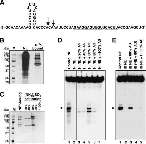
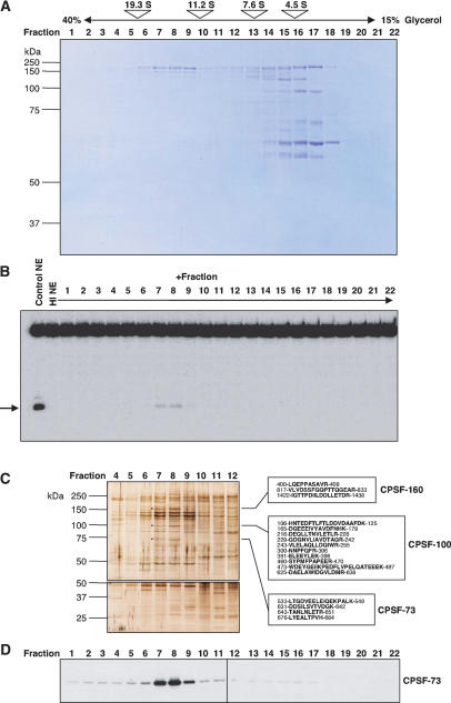
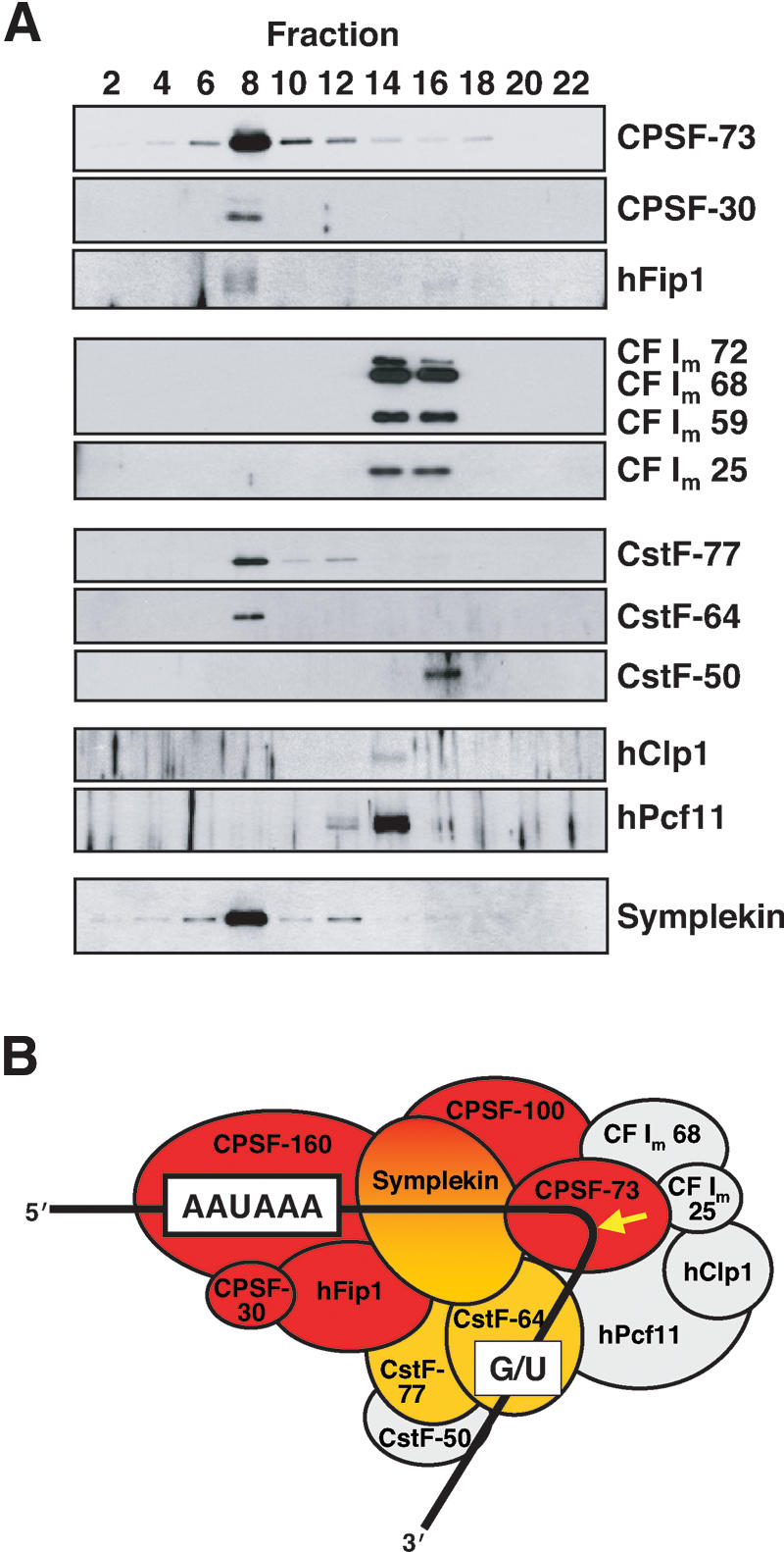
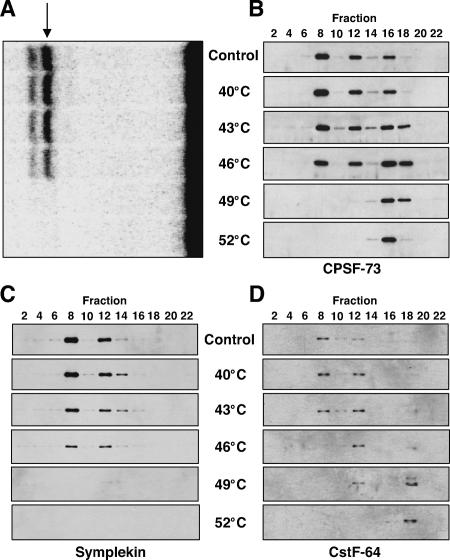
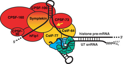
Most metazoan messenger RNAs encoding histones are cleaved, but not polyadenylated at their 3' ends. Processing in mammalian cell extracts requires the U7 small nuclear ribonucleoprotein (U7 snRNP) and an unidentified heat-labile factor (HLF). We describe the identification of a heat-sensitive protein complex whose integrity is required for histone pre-mRNA cleavage. It includes all five subunits of the cleavage and polyadenylation specificity factor (CPSF), two subunits of the cleavage stimulation factor (CstF), and symplekin. Reconstitution experiments reveal that symplekin, previously shown to be necessary for cytoplasmic poly(A) tail elongation and translational activation of mRNAs during Xenopus oocyte maturation, is the essential heat-labile component. Thus, a common molecular machinery contributes to the nuclear maturation of mRNAs both lacking and possessing poly(A), as well as to cytoplasmic poly(A) tail elongation.


Figures

Figure 1.
Figure 1.
HLF activity is retained on Ni2+-affinity resin. (A) The histone H4-12 pre-mRNA substrate used in the in vitro processing assays. The sequence that is complementary to the 5′-end of U7 snRNA is underlined. Large and small arrows indicate the alternative (major and minor) positions of endonucleolytic scission (Streit et al. 1993). (B) Coomassie brilliant blue-stained SDS-PAGE of the crude nuclear extract (NE) starting material (1/250th) and of the material eluted from Ni2+-NTA agarose (1/50th). Lane M shows molecular weight marker proteins. (C) Saturated (NH4)2SO4 was added progressively to the material eluted from the Ni2+ resin to achieve the indicated percent saturation values. Precipitated proteins were resuspended and visualized by Coomassie staining after separation on 10% SDS-PAGE. (D) Complementation of in vitro processing. HeLa nuclear extract (NE) was incubated at 50°C for 15 min to generate heat-inactivated extract (HI), which was used for processing of a uniformly labeled H4-12 pre-mRNA substrate either without supplementation (lane 2) or after addition of the indicated ammonium sulfate (AS) precipitates (lanes 3-7). Lane 1 shows processing in the extract before heat inactivation. The arrow indicates the upstream fragment (30 nt) produced by cleavage at the major site indicated in A; additional bands above and below are degradation products of the downstream fragment (36 nt). (E) The 60% ammonium sulfate fraction containing HLF activity (60% AS) is inactivated by heat treatment (15 min at 50°C). In vitro processing using a 5′-end labeled H4-12 pre-mRNA substrate. (Lane 1) Non-heat-treated extract. (Lane 2) Heat-inactivated extract. (Lane 3) Heat-inactivated extract complemented with the non-heat-treated 60% AS fraction. (Lane 4) A nontreated extract and the 60% AS fraction were mixed and incubated together at 50°C. (Lane 5) The extract and the 60% AS fraction were heat treated separately, then combined prior to addition of the processing substrate. The arrow indicates the upstream product resulting from cleavage at the major site; the faint band above results from processing at the minor position shown in A.
Figure 2.
Figure 2.
HLF activity sediments at 14.7S. (A) Protein profile of the 60% ammonium sulfate precipitate after 15%-40% glycerol gradient fractionation. Proteins were separated by 10% SDS-PAGE and stained with Coomassie. Above is shown the distribution of size markers in a parallel gradient: porcine thyroglobulin (19.3S, 669 kDa), bovine catalase (11.2S, 232 kDa), bovine lactate dehydrogenase (7.6S, 140 kDa), and bovine serum albumin (4.5S, 66 kDa). It should be noted that purified CPSF has a sedimentation coefficient of 11.5S (Bienroth et al. 1991; Murthy and Manley 1992), even though the predicted combined molecular weight of its subunits (excluding hFip1) is 357 kDa. (B) Complementation of in vitro processing by glycerol gradient fractions. Glycerol gradient fractions shown in A were mixed with heat-inactivated extract and incubated with a 5′-end labeled H4-12 substrate. The arrow indicates the major upstream cleavage product. We calculate (accounting for the dilution in the gradient) that fractions 7, 8, and 9 (14.7S fraction) combined contain slightly more HLF activity than the starting 60% ammonium sulfate precipitate. This indicates that there is no significant HLF stimulatory activity sedimenting with the bulk of the protein at ∼4.5S in our gradients. (C) SDS-PAGE (7% gel, top; 13% gel, bottom) of the fractions in the vicinity of HLF activity in the glycerol gradient shown in A was followed by silver staining. The boxes show tryptic peptide sequences identified by LC MS/MS analysis for the bands marked with black dots. Numbers indicate the amino acid positions within the sequences of the identified proteins. (D) Western blot analysis of all fractions from the glycerol gradient of A using anti-CPSF-73 antibody.
Figure 3.
Figure 3.
CPSF, CstF subunits, and symplekin cofractionate with HLF activity. (A) Western blots of fractions from a glycerol gradient separating the 60% ammonium sulfate precipitate, as in Figure 2. Only even-numbered fractions were analyzed for the presence of the proteins listed on the right. HLF activity peaked in fraction 8 (data not shown). (B) Diagram depicting proteins assigned to the cleavage and polyadenylation machinery, excluding poly(A) polymerase (Ryan et al. 2004). It has been previously observed that nuclear PAP does not copurify with either CPSF or CstF (Takagaki et al. 1989, and references therein). Consensus sequences and the site of cleavage (arrow) prior to polyadenylation are shown. HLF proteins concentrated in fraction 8, as identified by either mass spectrometry (Fig. 2C) or Western blotting (A) are colored red (CPSF) and orange (CstF). Proteins peaking elsewhere in the gradient are colored gray.
Figure 4.
Figure 4.
Heat destroys the integrity of the 14.7S HLF complex. (A) In vitro processing of histone pre-mRNA in HeLa nuclear extracts pretreated for 15 min at the indicated temperatures compared with a non-heat-treated extract (Control). The H4-12 substrate was internally 32P-labeled. The arrow indicates the upstream cleavage product. Additional bands above and below are degradation products of the downstream fragment. (B-D) The heat-treated nuclear extracts assayed in A, as well as a non-heat-treated extract (Control), were fractionated on 15%-40% glycerol gradients. Every other fraction was subjected to SDS-PAGE and immunoblotted with antibodies against CPSF-73, Symplekin, or CstF-64.
Figure 5.
Figure 5.
In vitro-translated symplekin restores processing activity in heat-inactivated extract. (Left) Western blots analyzing the presence of symplekin (top) or CPSF-73 (bottom) in equal volumes of non-heat-treated nuclear extract (lane 1), heat-inactivated extract (lane 2), symplekin cDNA-programmed TNT reaction (lane 3), or an unprogrammed TNT reaction (lane 4). Note that the symplekin band does not disappear immediately after heat treatment, suggesting that its heat-induced inactivation precedes its degradation during the lengthy centrifugation in the glycerol gradients. The right panel shows in vitro processing of an internally 32P-labeled H4-12 pre-mRNA substrate in control non-heat-inactivated nuclear extract (lane 1), heat-inactivated (HI) extract (lane 2), HI extract supplemented with a transcription/translation reaction (TNT) programmed with symplekin cDNA (lane 3), or HI extract supplemented with an unprogrammed TNT reaction (lane 4).
Figure 6.
Figure 6.
Model for histone pre-mRNA processing. Components of the HLF identified in this study are colored red and orange. The U7 snRNA is depicted base pairing with the histone pre-mRNA downstream element. We propose that the U7 snRNP orients the histone pre-mRNA for cleavage (arrow) by CPSF-73 through contacts between its Sm proteins and the CstF subunits in HLF (see text). Proteins of the Sm ring that are shared with spliceosomal snRNPs are shown in green; the U7-specific Lsm10 and Lsm11 proteins (Pillai et al. 2001, 2003) are shown in shades of blue.

Comment in

References

    1. Abbott J., Marzluff, W.F., and Gall, J.G. 1999. The stem-loop binding protein (SLBP1) is present in coiled bodies of the Xenopus germinal vesicle. Mol. Biol. Cell 10: 487-499. - PMC - PubMed
    1. Barabino S.M., Hübner, W., Jenny, A., Minvielle-Sebastia, L., and Keller, W. 1997. The 30-kD subunit of mammalian cleavage and polyadenylation specificity factor and its yeast homolog are RNA-binding zinc finger proteins. Genes & Dev. 11: 1703-1716. - PubMed
    1. Barnard D.C., Ryan, K., Manley, J.L., and Richter, J.D. 2004. Symplekin and xGLD-2 are required for CPEB-mediated cytoplasmic polyadenylation. Cell 119: 641-651. - PubMed
    1. Bienroth S., Wahle, E., Suter-Crazzolara, C., and Keller, W. 1991. Purification of the cleavage and polyadenylation factor involved in the 3′-processing of messenger RNA precursors. J. Biol. Chem. 266: 19768-19776. - PubMed
    1. Birchmeier C., Schümperli, D., Sconzo, G., and Birnstiel, M.L. 1984. 3′ editing of mRNAs: Sequence requirements and involvement of a 60-nucleotide RNA in maturation of histone mRNA precursors. Proc. Natl. Acad. Sci. 81: 1057-1061. - PMC - PubMed

Publication types