. 2005 Oct 15;19(20):2466-76.
doi: 10.1101/gad.1352105.

Replication of mitochondrial DNA occurs by strand displacement with alternative light-strand origins, not via a strand-coupled mechanism

Affiliations

Replication of mitochondrial DNA occurs by strand displacement with alternative light-strand origins, not via a strand-coupled mechanism

Timothy A Brown et al. Genes Dev. .

Abstract

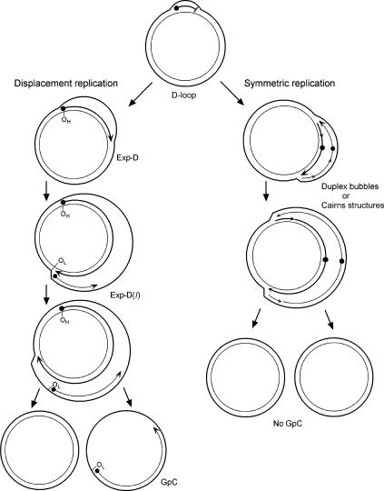
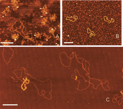
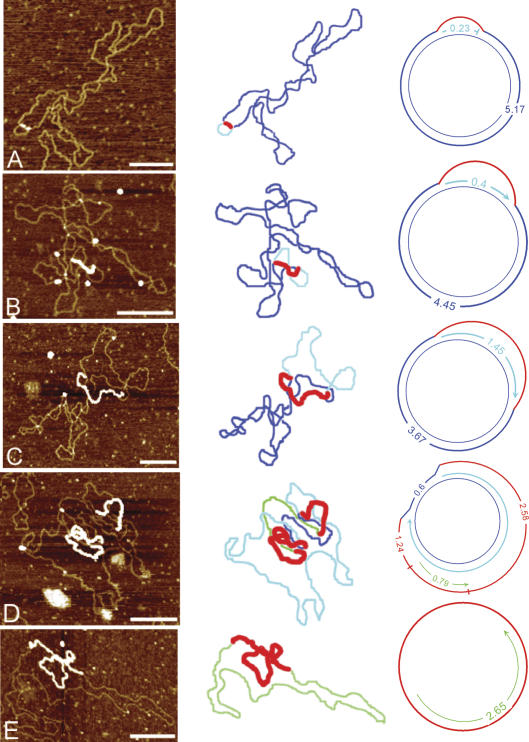
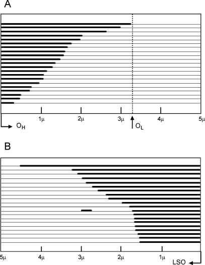
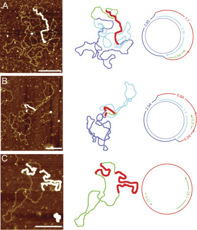
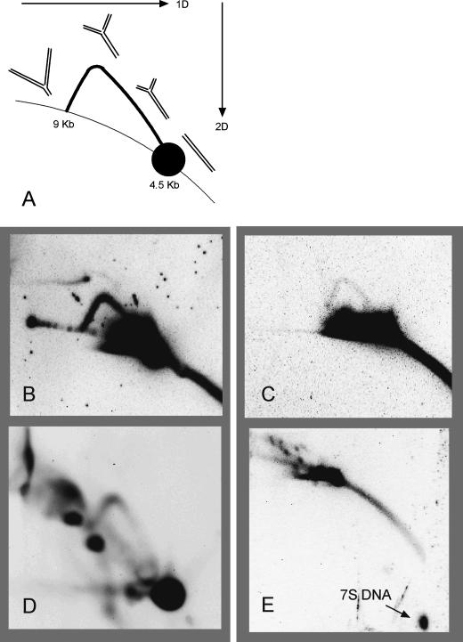
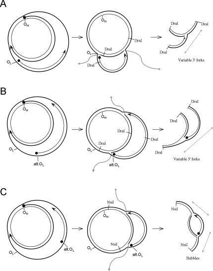
The established strand-displacement model for mammalian mitochondrial DNA (mtDNA) replication has recently been questioned in light of new data using two-dimensional (2D) agarose gel electrophoresis. It has been proposed that a synchronous, strand-coupled mode of replication occurs in tissues, thereby casting doubt on the general validity of the "orthodox," or strand-displacement model. We have examined mtDNA replicative intermediates from mouse liver using atomic force microscopy and 2D agarose gel electrophoresis in order to resolve this issue. The data provide evidence for only the orthodox, strand-displacement mode of replication and reveal the presence of additional, alternative origins of lagging light-strand mtDNA synthesis. The conditions used for 2D agarose gel analysis are favorable for branch migration of asymmetrically replicating nascent strands. These data reconcile the original displacement mode of replication with the data obtained from 2D gel analyses.


Figures

Figure 1.
Figure 1.
The asymmetric and strand-coupled models of mtDNA replication. Both models agree on the nature of the simple D-loop form of mtDNA. The displacement-model of replication is shown on the left and proceeds with single-stranded replication of the H-strand with further expansion and displacement of the D-loop. The intermediates are called expanded D-loops (Exp-D). This proceeds until the L-strand origin (OL) is exposed, with subsequent synthesis of the new L-strand in the opposite direction. Those intermediates are termed Exp-D(l). The asymmetry of strand synthesis leaves one segregated daughter molecule with an incompletely synthesized L-strand, called a gapped circle (GpC). The strand-coupled or synchronous model of replication is shown on the right. In this model, there is thought to be a zone of replication initiation within a broad area beyond the simple D-loop. Within this zone, both strands are synthesized bidirectionally as the double-stranded replication forks proceed through the length of the mtDNA. Exp-D, Exp-D(l), and GpC forms are excluded by this mode of replication.
Figure 2.
Figure 2.
AFM images of M13 phage and mtDNA. M13 phage genomes before (A) and after (B) binding to SSB. (C) Two mtDNA molecules with single-stranded regions coated with SSB. Bars, 0.25 μm.
Figure 3.
Figure 3.
Replicative intermediates of mtDNA seen by AFM. The first column displays the AFM images, the second column outlines the contour, and the third column diagrams the intermediate and shows the strand contour lengths. Blue lines represent the parental strands, red lines represent the compacted ssDNA coated with SSB protein, aqua lines represent nascent H-strand synthesis, and green lines represent nascent L-strand synthesis. (A) Simple D-loop. (B) Short Exp-D. (C) Large Exp-D. (D) Exp-D with L-strand (Exp-D[l]). (E) GpC molecule with partial synthesis of the L-strand. Bars, 0.25 μm.
Figure 4.
Figure 4.
Length distribution of newly synthesized nascent-strand intermediates. Individual mtDNA molecules are shown in linear form. Thick lines represent new complementary strand synthesis, and parental strands are shown as thin lines. (A) The lengths of the newly synthesized heavy strands measured from Exp-D molecules. All Exp-D in this analysis occur within the boundaries of OH and OL. (B) The lengths of nascent L-strands measured from GpC molecules. The position of OH cannot be determined from the GpC forms and is therefore not shown. The L-strand origin (LSO) is displayed to the far right.
Figure 5.
Figure 5.
Replicating mtDNA with alternate L-strand origins. The first column displays the AFM images, the second column outlines the contour, and the third column diagrams the intermediate and shows the strand contour lengths. Blue lines represent the parental strands, red lines represent the compacted ssDNA coated with SSB protein, aqua lines represent the nascent H-strand synthesis, and green lines represent nascent L-strand synthesis. Although the third column shows the molecules in one particular orientation relative to OH, the opposite orientation is also possible, with OH located at the other fork. Bars, 0.25 μm.
Figure 6.
Figure 6.
Neutral–neutral 2D agarose gel analysis of mouse mtDNA. (A) Diagram of a y-arc. The orientations of the first and second dimensions are shown with arrows. The formation of the y-arc is described in the text. In B and C, the 4.5-kb DraI fragment was probed with a portion of the ATPase 6 gene (nucleotides 8032–8497) and was treated with S1 nuclease. DNA in D was digested with BglI and probed with a fragment of the ND4 gene (nucleotides 10577–11193). DNA in E was probed for the 7S DNA strand, corresponding to the D-loop region (nucleotides 16027–15510). DNA in D and E was not treated with S1 nuclease. (B) 300 ng liver mtDNA. (C) 500 ng LA9 mtDNA. (D) 500 ng liver mtDNA. (E) 150 ng liver mtDNA.
Figure 7.
Figure 7.
Formation of y- and bubble-arc intermediates through alternate L-strand origins and branch migration of displacement replication strands. Branch-migrated arms of nascent strands are shown in gray. (A) Variable fork formation defined by the 3′ end of the L-strand. (B) Variable fork formation defined by the 5′ end of the L-strand. Variable formation of this end is dependent upon alternative L-strand initiation. (C) Variable fork formation at both ends to yield bubble-type intermediates.

References

    1. Aloni Y. and Attardi, G. 1972. Expression of the mitochondrial genome in HeLa cells XI. Isolation and characterization of transcription complexes of mitochondrial DNA. J. Mol. Biol. 70: 363-373. - PubMed
    1. Bell L. and Byers, B. 1983. Separation of branched from linear DNA by two-dimensional gel electrophoresis. Anal. Biochem. 130: 527-535. - PubMed
    1. Berk A.J. and Clayton, D.A. 1974. Mechanism of mitochondrial DNA replication in mouse L-cells: Asynchronous replication of strands, segregation of circular daughter molecules, aspects of topology and turnover of an initiation sequence. J. Mol. Biol. 86: 801-824. - PubMed
    1. Bogenhagen D.F. and Clayton, D.A. 2003a. The mitochondrial DNA replication bubble has not burst. Trends Biochem. Sci. 28: 357-360. - PubMed
    1. ____. 2003b. Concluding remarks: The mitochondrial DNA replication bubble has not burst. Trends Biochem. Sci. 28: 404-405. - PubMed

MeSH terms

Substances

LinkOut - more resources