. 2005 Oct 15;19(20):2501-15.
doi: 10.1101/gad.344505.

The microtubule plus-end-tracking protein CLIP-170 associates with the spermatid manchette and is essential for spermatogenesis

Affiliations

The microtubule plus-end-tracking protein CLIP-170 associates with the spermatid manchette and is essential for spermatogenesis

Anna Akhmanova et al. Genes Dev. .

Abstract

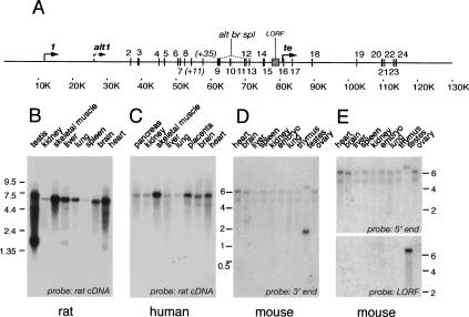
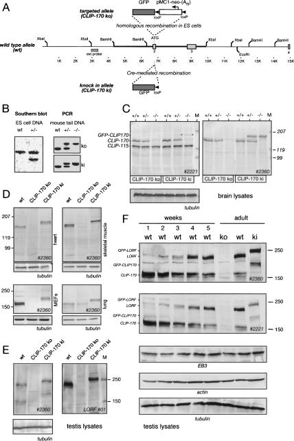
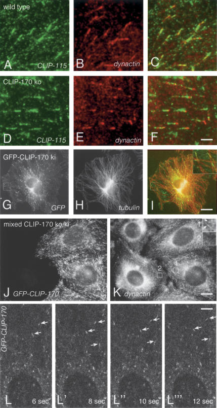
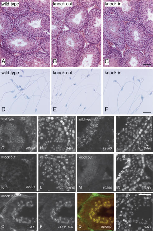
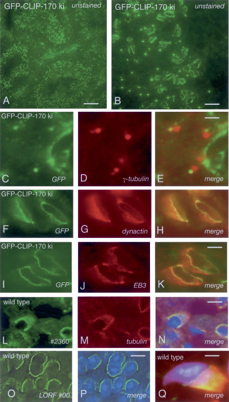
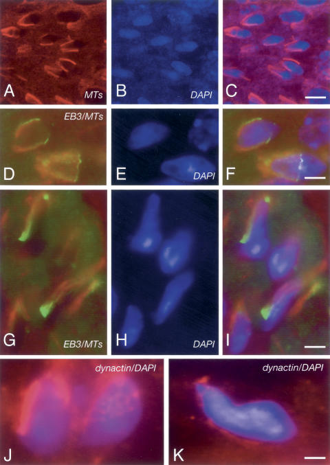
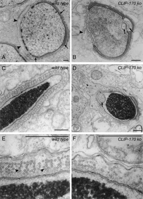
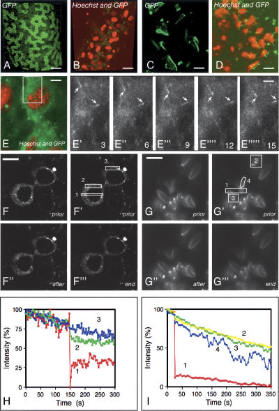
CLIP-170 is a microtubule "plus-end-tracking protein" implicated in the control of microtubule dynamics, dynactin localization, and the linking of endosomes to microtubules. To investigate the function of mouse CLIP-170, we generated CLIP-170 knockout and GFP-CLIP-170 knock-in alleles. Residual CLIP-170 is detected in lungs and embryos of homozygous CLIP-170 knockout mice, but not in other tissues and cell types, indicating that we have generated a hypomorphic mutant. Homozygous CLIP-170 knockout mice are viable and appear normal. However, male knockout mice are subfertile and produce sperm with abnormal heads. Using the knock-in mice, we followed GFP-CLIP-170 expression and behavior in dissected, live testis tubules. We detect plus-end-tracking GFP-CLIP-170 in spermatogonia. As spermatogenesis proceeds, GFP-CLIP-170 expression increases and the fusion protein strongly marks syncytia of differentiated spermatogonia and early prophase spermatocytes. Subsequently GFP-CLIP-170 levels drop, but during spermiogenesis (post-meiotic development), GFP-CLIP-170 accumulates again and is present on spermatid manchettes and centrosomes. Bleaching studies show that, as spermatogenesis progresses, GFP-CLIP-170 converts from a mobile plus-end-tracking protein to a relatively immobile protein. We propose that CLIP-170 has a structural function in the male germline, in particular in spermatid differentiation and sperm head shaping.


Figures

Figure 1.
Figure 1.
Genomic organization and expression of the gene encoding CLIP-170. (A) Genomic organization of the gene encoding CLIP-170. The position of common (1-24) and alternatively spliced (+11, +35, LORF) exons and transcription initiation sites (1, alt1, te) is indicated. (Alt br spl) Alternative splice event generating the brain-specific mRNA. Scale in kilobase pairs (K). (B-E) CLIP-170 mRNA expression. Northern blots from rat, human, and mouse were hybridized with the indicated probes. Size markers are indicated (kilobases).
Figure 2.
Figure 2.
Generation of CLIP-170 KO and GFP-CLIP-170 KI in alleles. (A) Targeting strategy of CLIP-170 KO and GFP-CLIP-170 KI alleles. A segment of the murine Rsn gene is shown, with the ATG-containing exon and diagnostic restriction enzyme sites. The 5′-end and 3′-end homologous arms of the targeting construct are indicated by thick lines. The insertion cassette in the targeting construct is shown above the Rsn gene and contains GFP, loxP sites (arrowheads), and a neomycin resistance (neo) gene, driven by the pMC1 promoter and containing a polyadenylation sequence (An). The neo gene is transcribed antisense (see arrow) to the Rsn gene. Homologous recombination generates a CLIP-170 KO allele. Cre-mediated excision at the targeted Rsn allele leaves GFP and one loxP site. As these are inframe with CLIP-170-encoding sequences, the GFP-CLIP-170 KI allele is generated. (B) Southern blot and PCR analysis. The Southern blot demonstrates the presence of a homologously targeted CLIP-170 KO allele in ES cells. The PCR analysis with three primers allows detection of the wild-type and CLIP-170 KO or GFP-CLIP-170 KI alleles. (C-F) Western blots of total protein lysates from brain (C), heart, skeletal muscle, passage 30 MEFs, and lung (D) or testis (E,F), incubated with antibodies recognizing both CLIP-115 and CLIP-170 (#2221), CLIP-170 only (#2360), or the LORF-containing CLIP-170 isoform (LORF#01), tubulin, EB3, and actin. (C) The #2221 antibodies recognize GFP-CLIP-170 less well (asterisks in lanes). For each tissue, equal amounts of protein were loaded in each lane. In F, testis lysates were prepared from mice ranging in age from 1 wk to adult. Size markers (M) are indicated (kilodaltons).
Figure 3.
Figure 3.
Cellular analysis of the CLIP-170 deletion and GFP-CLIP-170 insertion. (A-F) CLIP-115 and dynactin distribution in wild-type (A-C) and CLIP-170 KO MEFs (D-F). CLIP-115 (green) and dynactin (red) were detected with antibodies #2238 and antibodies against dynamitin, respectively. Both CLIP-115 and dynactin localize at the ends of MTs, but dynactin signal is diminished in KO MEFs. Bar, 2.5 μm. (G-I) GFP-CLIP-170 distribution in KI MEFs. Direct GFP fluorescence (green) was observed in combination with anti-tubulin staining (red). Enlarged MT plus-ends are shown in the inset. Bar, 10 μm. (J,K) Dynactin distribution in keratinocytes. A mixed culture of CLIP-170 KO and GFP-CLIP-170 KI keratinocytes was made by transfecting a KO culture with Cre recombinase. GFP-CLIP-170 is shown in J (direct fluorescence) and dynactin in K. Notice the decreased dynactin staining at MT ends in KO cells (see also insets in K). Bar, 13 μm. (L-L′′′) Behavior of GFP-CLIP-170 in cultured glial cells, derived from the GFP-CLIP-170 KI mice, analyzed by time-lapse imaging. The movie of this analysis is shown as Supplementary Movie 1. L-L′′′ represent parts of the images acquired after 6, 8, 10, and 12 sec. The arrows indicate three examples of moving GFP-CLIP-170 dashes. Bar, 3 μm.
Figure 4.
Figure 4.
Testicular analysis of the CLIP-170 deletion and GFP-CLIP-170 insertion. (A-C) H/E-stained 7-μm sections of the testes from wild-type, CLIP-170 KO, and GFP-CLIP-170 KI mice. Bar, 50 μm. (D-F) Sperm preparations, isolated from the epididymis of wild-type, CLIP-170 KO, and GFP-CLIP-170 KI mice. Bar, 10 μm. (G-R) Immunofluorescence analysis. Wild-type (G-J), CLIP-170 KO (K-N), and GFP-CLIP-170 KI (O-R) paraffin-embedded sections were incubated with the indicated antibodies and counterstained with DAPI to visualize nuclei. In the overlay in Q, anti-GFP staining is shown in green, and LORF#00 in red. Bar, 40 μm.
Figure 5.
Figure 5.
CLIP-170 and GFP-CLIP-170 distribution in wild-type and KI testis. (A,B) Unstained cryosections from GFP-CLIP-170 KI mice. Bars: A, 40 μm; B, 8 μm. (C-K) Localization of GFP-CLIP-170 to the centrosomes and the spermatid manchette. Testis cryosections from GFP-CLIP-170 KI mice were incubated with antibodies against γ-tubulin (C-E), dynactin subunit p150Glued (F-H), or EB3 (I-K). GFP was detected by its direct fluorescence. Bars: E, 1.5 μm; K, 2.5 μm. (L-Q) Paraffin-embedded testis sections from wild-type mice were incubated with #2360, or LORF antibodies (green), β-tubulin antisera (red), and counterstained with DAPI (blue). In L-N, images were acquired with a confocal microscope and deconvolved. Notice how CLIP-170 colocalizes with tubulin. In O and P, images were acquired with an epifluorescent microscope and deconvolved. A 3D representation of the LORF-CLIP-170 distribution is shown in Supplementary Movie 2. In Q, the image was acquired with an epifluorescent microscope and is only shown merged. Bars: N,P, 5 μm; Q, 1.4 μm.
Figure 6.
Figure 6.
Immunofluorescent analysis of CLIP-170 KO testis. (A-I) Paraffin-embedded testis sections from CLIP-170 KO mice were incubated with antibodies against β-tubulin (red) (A-C), or β-tubulin (red) and EB3 (green) (D-I) and counterstained with DAPI (blue). A 3D representation of EB3 and MT distribution is shown in Supplementary Movie 3. Bars: C, 8 μm; F, 2.5 μm; I, 4 μm. (J,K) Cryosections of the testis from CLIP-170 KO mice were incubated with antibodies against dynactin subunit p150Glued (red) and counterstained with DAPI (blue). Bar, 1 μm.
Figure 7.
Figure 7.
Ultrastructural morphology of the testis of wild-type and CLIP-170 KO mice. (A) Step 8 spermatid from a wild-type mouse with manchette MTs (indicated by arrowheads) located in close apposition to the nucleus in the area not occupied by the acrosome (indicated by arrows). (B) Step 8 spermatid from a CLIP-170 KO mouse. Some manchette MTs appear to be properly positioned (arrowhead). However, they do not completely cover the area of the nucleus that is free of the acrosome. Moreover, bundles of MTs show ectopic expression and are positioned within the nucleus (arrows). (C,D) Transverse sections from wild-type (C) or CLIP-170 KO (D) step 14 spermatids, showing portions of the manchette and nucleus. In the wild-type spermatid, the nucleus is surrounded by highly regularly organized MT bundles, which form a well-developed manchette. The CLIP-170 KO nucleus is almost round, and the manchette shows ectopic expression with MTs detached from the nucleus (arrowhead) and within the nucleus (arrow). (E) Higher magnification of a part of the wild-type manchette shown in C. The cross-sectioned microtubules can be seen arranged in a regular pattern, with prominent cross-bridges (arrowheads). (F) High magnification of a part of the KO manchette shown in D. Cross-sectioned MTs in KO mice display a highly irregular arrangement. Not all MTs are truly cross-sectioned, indicating that they do not run in parallel arrays. Bars, 0.5 μm.
Figure 8.
Figure 8.
Behavior of GFP-CLIP-170 in live testis tubules. Testis tubules were dissected from GFP-CLIP-170 KI mice (in some cases treated with Hoechst) and analyzed with a confocal/multiphoton microscope. (A) Low-magnification view of GFP-CLIP-170 distribution. The panel shows an image from Supplementary Movie 4. Notice the accumulation of GFP-CLIP-170 in pre-leptotene spermatocytes. Bar, 30 μm. (B) Low-magnification view of GFP-CLIP-170 and Hoechst distribution. The panel shows an image from Supplementary Movie 5. Notice the accumulation of GFP-CLIP-170 in type B spermatogonia and the absence of signal in Sertoli cells. Bar, 25 μm. (C) High-magnification view of GFP-CLIP-170 distribution in elongating spermatids. Analysis was as in A. Notice the accumulation of GFP-CLIP-170 on manchettes, the manchette ring, and on centrosomes. Bar, 5 μm. (D) High-magnification view of GFP-CLIP-170 and Hoechst distribution in elongating spermatids. Analysis was as in B. This panel shows one plane from Supplementary Movie 7. Notice the accumulation of GFP-CLIP-170 in elongating but not in the round spermatids. Bar, 6 μm. (E-E′′′) Time-lapse analysis of GFP-CLIP-170. Analysis was as in B. In E, a combined GFP and Hoechst image is shown to identify the germ cell as a differentiated type A spermatogonium. Subsequent time-lapse analysis was done with the 488-nm laser only and is shown in Supplementary Movie 8. E′-E′′′ show part of the images (see rectangle in E) acquired after 3, 6, 9, 12, and 15 sec, respectively. Arrows indicate GFP-CLIP-170-positive dashes. Bar, 6 μm. (F-I) FRAP/FLIP analysis of GFP-CLIP-170 behavior. Live images of a portion of a testis tubule were taken, and after a number of frames, the rectangular regions indicated with the number 1 were bleached and time-lapse analysis was resumed. In F and H, undifferentiated spermatogonia (A-paired) were analyzed, whereas in G and I, elongating spermatids were analyzed. H and I show the relative fluorescent intensity (percentage) of the indicated regions of interest (ROIs) during the experiment. Whereas GFP-CLIP-170 is largely mobile in spermatogonia, it is largely immobile in spermatids. Bars: F, 9 μm; G, 6 μm.

Similar articles

Cited by

References

    1. Akhmanova A. and Hoogenraad, C.C. 2005. Microtubule plus-end-tracking proteins: Mechanisms and functions. Curr. Opin. Cell Biol. 17: 47-54. - PubMed
    1. Akhmanova A., Hoogenraad, C.C., Drabek, K., Stepanova, T., Dortland, B., Verkerk, T., Vermeulen, W., Burgering, B.M., De Zeeuw, C.I., Grosveld, F., et al. 2001. Clasps are CLIP-115 and -170 associating proteins involved in the regional regulation of microtubule dynamics in motile fibroblasts. Cell 104: 923-935. - PubMed
    1. Arnal I., Heichette, C., Diamantopoulos, G.S., and Chretien, D. 2004. CLIP-170/tubulin-curved oligomers coassemble at microtubule ends and promote rescues. Curr. Biol. 14: 2086-2095. - PubMed
    1. Bilbe G., Delabie, J., Bruggen, J., Richener, H., Asselbergs, F.A., Cerletti, N., Sorg, C., Odink, K., Tarcsay, L., Wiesendanger, W., et al. 1992. Restin: A novel intermediate filament-associated protein highly expressed in the Reed-Sternberg cells of Hodgkin's disease. EMBO J. 11: 2103-2113. - PMC - PubMed
    1. Brunner D. and Nurse, P. 2000. CLIP170-like tip1p spatially organizes microtubular dynamics in fission yeast. Cell 102: 695-704. - PubMed

Publication types

MeSH terms

Substances

LinkOut - more resources