Complete and persistent phenotypic correction of phenylketonuria in mice by site-specific genome integration of murine phenylalanine hydroxylase cDNA
- PMID: 16230623
- PMCID: PMC1266087
- DOI: 10.1073/pnas.0503877102
Complete and persistent phenotypic correction of phenylketonuria in mice by site-specific genome integration of murine phenylalanine hydroxylase cDNA
Retraction in
-
Retraction for Chen et al., Complete and persistent phenotypic correction of phenylketonuria in mice by site-specific genome integration of murine phenylalanine hydroxylase cDNA.Proc Natl Acad Sci U S A. 2010 Aug 10;107(32):14514. doi: 10.1073/pnas.1011158107. Epub 2010 Jul 9. Proc Natl Acad Sci U S A. 2010. PMID: 20622151 Free PMC article. No abstract available.
Abstract
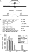
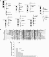
We explored the potential of using a bacteriophage integrase system to achieve site-specific genome integration of murine phenylalanine hydroxylase cDNA in the livers of phenylketonuric (PKU) mice. The phiBT1 phage integrase is an enzyme that catalyses the efficient recombination between unique sequences in the phage and bacterial genomes, leading to the site-specific integration of the former into the latter in a unidirectional manner. Here we showed that this phage integrase functions efficiently in mouse cells, and several naturally occurring pseudo-attP sites located in the intergenic regions of the mouse genome have been identified and molecularly characterized. We further demonstrated that the addition of nuclear localization signal sequences to the C terminus of the phage integrase enhanced the efficiency for transgene integration into the mouse genome. Using this phage integration system, we delivered mouse phenylalanine hydroxylase cDNA to the livers of PKU mice by hydrodynamic injection of plasmid DNA and showed that the severity of the hyperphenylalaninemic phenotype in the treated mice decreased significantly. After three applications, serum phenylalanine levels in all treated PKU mice were reduced to the normal range and remained stable thereafter. Their fur color also changed from gray to black, indicating the reconstitution of melanin biosynthesis as a result of available tyrosine derived from reconstituted phenylalanine hydroxylation in the liver. Thus, the phiBT1 bacteriophage integrase represents an effective site-specific genome integration system in mammalian cells and can be of great value in DNA-mediated gene therapy for a multitude of genetic disorders.
Figures


Comment in
-
Findings of research misconduct.NIH Guide Grants Contracts (Bethesda). 2014 May 23:NOT-OD-14-098. NIH Guide Grants Contracts (Bethesda). 2014. PMID: 24864370 Free PMC article. No abstract available.
-
Findings of Research Misconduct.Fed Regist. 2014 Apr 25;79(80):22973-22975. Fed Regist. 2014. PMID: 27737255 Free PMC article. No abstract available.
Similar articles
-
Metabolic basis of sexual dimorphism in PKU mice after genome-targeted PAH gene therapy.Mol Ther. 2007 Jun;15(6):1079-85. doi: 10.1038/sj.mt.6300137. Epub 2007 Apr 3. Mol Ther. 2007. Retraction in: Mol Ther. 2010 Dec;18(12):2190. doi: 10.1038/mt.2010.198. PMID: 17406346 Retracted.
-
Site-specific transgene integration in the human genome catalyzed by phiBT1 phage integrase.Hum Gene Ther. 2008 Feb;19(2):143-51. doi: 10.1089/hum.2007.110. Hum Gene Ther. 2008. Retraction in: Hum Gene Ther. 2010 Aug;21(8):1036. doi: 10.1089/hum.2010.1513. PMID: 18067406 Retracted.
-
Correction in female PKU mice by repeated administration of mPAH cDNA using phiBT1 integration system.Mol Ther. 2007 Oct;15(10):1789-95. doi: 10.1038/sj.mt.6300257. Epub 2007 Jul 17. Mol Ther. 2007. Retraction in: Mol Ther. 2010 Dec;18(12):2191. doi: 10.1038/mt.2010.199. PMID: 17637719 Retracted.
-
Gene therapy for phenylketonuria.Acta Paediatr Suppl. 1994 Dec;407:124-9. doi: 10.1111/j.1651-2227.1994.tb13471.x. Acta Paediatr Suppl. 1994. PMID: 7766948 Review.
-
State-of-the-Art 2019 on Gene Therapy for Phenylketonuria.Hum Gene Ther. 2019 Oct;30(10):1274-1283. doi: 10.1089/hum.2019.111. Epub 2019 Sep 9. Hum Gene Ther. 2019. PMID: 31364419 Free PMC article. Review.
Cited by
-
Controlled and localized genetic manipulation in the brain.J Cell Mol Med. 2006 Apr-Jun;10(2):333-52. doi: 10.1111/j.1582-4934.2006.tb00403.x. J Cell Mol Med. 2006. PMID: 16796803 Free PMC article. Review.
-
Intracellular gene transfer in rats by tail vein injection of plasmid DNA.AAPS J. 2010 Dec;12(4):692-8. doi: 10.1208/s12248-010-9231-z. Epub 2010 Sep 22. AAPS J. 2010. PMID: 20859713 Free PMC article.
-
Identification of pharmacological chaperones as potential therapeutic agents to treat phenylketonuria.J Clin Invest. 2008 Aug;118(8):2858-67. doi: 10.1172/JCI34355. J Clin Invest. 2008. PMID: 18596920 Free PMC article.
-
Preferential delivery of the Sleeping Beauty transposon system to livers of mice by hydrodynamic injection.Nat Protoc. 2007;2(12):3153-65. doi: 10.1038/nprot.2007.471. Nat Protoc. 2007. PMID: 18079715 Free PMC article.
-
Predicting preferential DNA vector insertion sites: implications for functional genomics and gene therapy.Genome Biol. 2007;8 Suppl 1(Suppl 1):S12. doi: 10.1186/gb-2007-8-s1-s12. Genome Biol. 2007. PMID: 18047689 Free PMC article. Review.
References
Publication types
MeSH terms
Substances
LinkOut - more resources
Full Text Sources
Medical
Molecular Biology Databases

