. 2005 Oct 25;102(43):15593-8.
doi: 10.1073/pnas.0507865102. Epub 2005 Oct 17.

Insulin-like growth factors 1 and 2 induce lymphangiogenesis in vivo

Affiliations

Insulin-like growth factors 1 and 2 induce lymphangiogenesis in vivo

Meit Björndahl et al. Proc Natl Acad Sci U S A. .

Abstract

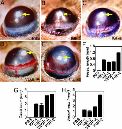
Lymphangiogenesis is an important process that contributes to the spread of cancer. Here we show that insulin-like growth factors 1 (IGF-1) and 2 (IGF-2) induce lymphangiogenesis in vivo. In a mouse cornea assay, IGF-1 and IGF-2 induce lymphangiogenesis as detected with LYVE-1, a specific marker for lymphatic endothelium. Interestingly, IGF-1-induced lymphangiogenesis could not be blocked by a soluble vascular endothelial growth factor receptor 3, suggesting that the vascular endothelial growth factor receptor 3-signaling pathway is not required for IGF-induced lymphangiogenesis. In vitro, IGF-1 and IGF-2 significantly stimulated proliferation and migration of primary lymphatic endothelial cells. IGF-1 and IGF-2 induced phosphorylation of intracellular signaling components, such as Akt, Src, and extracellular signal-regulated kinase in lymphatic endothelial cells. Immunohistochemistry, RT-PCR, and Affymetrix GeneChip microarray analysis showed that the receptors for IGFs are present in lymphatic endothelium. Together, our findings suggest that IGFs might act as direct lymphangiogenic factors, although any indirect roles in the induction of lymphangiogenesis cannot be excluded. Because members of the IGF ligand and receptor families are widely expressed in various types of solid tumors, our findings suggest that these factors are likely to contribute to lymphatic metastasis.


Figures

Fig. 1.
Fig. 1.
Stimulation of hemangiogenesis. (BE) Mouse corneas were implanted with 1 μg of IGF-1 (B), 0.5 μg of IGF-2 (C), 160 ng of VEGF-A165 (D), or 80 ng of FGF-2 (E). At day 5 after implantation, corneal neovascularization was photographed. (A) Slow-release polymer containing PBS without growth factors was used as a negative control. Arrows in AE point to the implanted pellets. (FH) Quantification of vessel length (F), clock-hour (G), and neovascularization area (H).
Fig. 2.
Fig. 2.
Stimulation of lymphangiogenesis. The IGF-1-stimulated (AD), IGF-2-stimulated (EH), or FGF-2-stimulated (IL) mouse corneas were removed at day 14 after growth factor implantation. (A, B, E, F, I, and J) The corneal tissues were stained with an anti-CD31 (A, E, and I) and an anti-LYVE-1 (B, F, and J) antibody. CD31-positive signals are in red, and LYVE-1-positive signals are in blue. (C, D, G, H, K, and L) The superimposed pictures of A and B, E and F, and I and J, respectively. (D, H, and L) Amplified images of boxed areas in C, G, and K. White arrows point to lymphatic vessels, and yellow arrows point to blood vessels. (Scale bar, 100 μm.)
Fig. 3.
Fig. 3.
Failure to inhibit IGF-1-induced corneal angiogenesis or lymphangiogenesis by soluble VEGFR-3. (AF) IGF-1-implanted (AC) or IGF-1/sVEGFR-3-implanted (DF) sections were double-stained with an anti-CD31 antibody (A and D, red) and an anti-LYVE-1 (B and E, blue) antibody. (G and H) Quantification of CD31-positive (G) and LYVE-1-positive (H) signals (n = 5–7 optical fields). (Scale bar, 100 μm.)
Fig. 4.
Fig. 4.
Expression of IGFRs of isolated LECs. (A and C) Isolated human LECs were stained with an anti-IGF-1R antibody. (D and F) Nonimmune rabbit IgG was used to stain the same LECs as a negative control. (B, C, E, and F) DAPI was used to stain cell nuclei. (G and H) Mouse (G) and human (H) LECs were used to prepare mRNA and cDNAs for RT-PCR amplification of IGF-1R and IGF-2R.
Fig. 5.
Fig. 5.
Proliferation, migration, and intracellular signaling pathway activation of LECs stimulated with IGFs. (AD) Mouse (A) and human (B, C, and D) LECs were used to study cell migration (AC) and cell proliferation (D). (AC) For analysis of chemotaxis, cells were stimulated with increasing concentrations of IGF-1 or IGF-2 or with 100 ng/ml IGF-1 or IGF-2, together with 500 ng/ml sR3. (D) For analysis of proliferation, cells were treated with 100 ng/ml IGF-1 or IGF-2. *, P < 0.05; **, P < 0.01; ***, P < 0.001. (E and F) Phosphorylation of the intracellular-signaling proteins ERK, Akt, and Src was analyzed in human LECs stimulated with 100 ng/ml IGF-1 (E) or IGF-2 (F) for different time periods.

References

    1. Fidler, I. J. (2003) Nat. Rev. Cancer 3, 453–458. - PubMed
    1. Beasley, N. J., Prevo, R., Banerji, S., Leek, R. D., Moore, J., van Trappen, P., Cox, G., Harris, A. L. & Jackson, D. G. (2002) Cancer Res. 62, 1315–1320. - PubMed
    1. Dadras, S. S., Lange-Asschenfeldt, B., Velasco, P., Nguyen, L., Vora, A., Muzikansky, A., Jahnke, K., Hauschild, A., Hirakawa, S., Mihm, M. C. & Detmar, M. (2005) Mod. Pathol. 18, 1232–1242. - PubMed
    1. Alitalo, K. & Carmeliet, P. (2002) Cancer Cell 1, 219–227. - PubMed
    1. Cao, R., Bjorndahl, M. A., Religa, P., Clasper, S., Garvin, S., Galter, D., Meister, B., Ikomi, F., Tritsaris, K., Dissing, S., et al. (2004) Cancer Cell 6, 333–345. - PubMed

Publication types

MeSH terms

Substances

LinkOut - more resources