. 2005 Oct 25;102(43):15563-8.
doi: 10.1073/pnas.0507835102. Epub 2005 Oct 17.

Human decidual NK cells form immature activating synapses and are not cytotoxic

Affiliations

Human decidual NK cells form immature activating synapses and are not cytotoxic

Hernan D Kopcow et al. Proc Natl Acad Sci U S A. .

Abstract

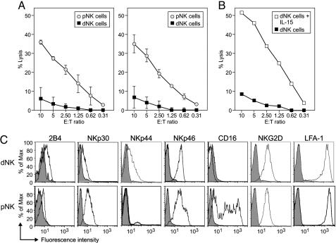
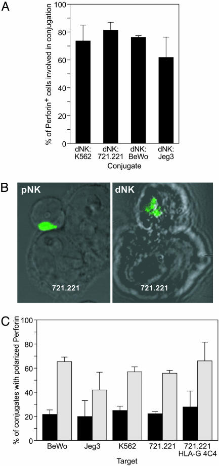
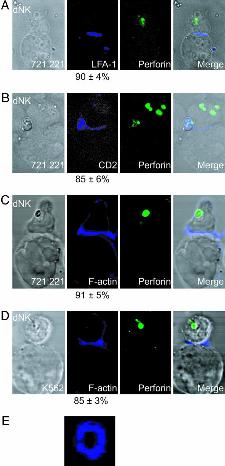
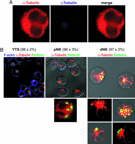
In early pregnancy invading fetal trophoblasts encounter abundant maternal decidual natural killer cells (dNK). dNK express perforin, granzymes A and B and the activating receptors NKp30, NKp44, NKp46, NKG2D, and 2B4 as well as LFA-1. Even though they are granular and express the essential molecules required for lysis, fresh dNK displayed very reduced lytic activity on classical MHC I negative targets K562 and 721.221, approximately 15% of that of peripheral NK cells. dNK formed conjugates and activating immune synapses with 721.221 and K562 cells in which CD2, LFA-1 and actin were polarized toward the contact site. However, in contrast to peripheral NK cells, they failed to polarize their microtubule organizing centers and perforin-containing granules to the synapse, accounting for their lack of cytotoxicity.


Figures

Fig. 1.
Fig. 1.
Freshly isolated dNK fail to lyse classical NK cell targets, although they express activating receptors. (A) Chromium release assays evaluating the lytic activity of dNK (filled squares) and pNK (open circles) cells on the classical NK cell target cell lines 721.221 (Left) and K562 (Right) (average of four independent experiments). (B) Cytotoxic activity of dNK (filled squares) and dNK from the same preparation incubated for 36 h with IL-15 (12 ng/ml) in RPMI medium 1640 plus 10% FCS (open squares) on 721.221 targets (one representative experiment of three). (C) FACS analysis of the expression of activating receptors by dNK and pNK (IgG isotype controls in gray).
Fig. 2.
Fig. 2.
dNK form conjugates but fail to polarize perforin granules toward the target cell contact site. (A) Conjugate formation by dNK visualized under the microscope. dNK were identified as perforin-positive cells. Bars represent the percentage of perforin positive cells (n ≥ 70 per slide) involved in conjugates with K562, 721.221, BeWo, or Jeg3 targets (average of three independent experiments). (B) Confocal micrographs showing a typical pNK-721.221 conjugate with polarized perforin granules (Left) and a typical dNK-721.221 conjugate with unpolarized perforin granules (Right). Perforin granules are visualized in green. (C) Perforin polarization in dNK (black bars) or pNK (gray bars) conjugated with different target cells as visualized by confocal microscopy. The percentage of conjugates with perforin containing granules polarized toward the target cell is shown (average of three independent experiments).
Fig. 3.
Fig. 3.
dNK form immature activating immune synapses. LFA-1, CD2, and F-actin, but not perforin, polarize to the target cell contact site in dNK. dNKs are shown conjugated with 721.221 cells (AC) and K562 cells (D) in four different conjugates. Differential interference contrast (DIC) images are shown at Left. LFA-1, CD2, or F-actin (blue) are shown in Center Left. Numbers indicate the percentage of conjugates with polarized LFA-1, CD2, or F-actin respectively. Perforin granules (green) are shown in Center Right. Merged overlays of all fluorescent and DIC channels at Right. (E) F-actin forming a pSMAC ring at the NKIS of a dNK-K562 conjugate.
Fig. 4.
Fig. 4.
Perforin containing granules are clustered around the MTOC in resting NK cells. (A) Confocal image of a YTS cell showing the microtubule cytoskeleton stained with anti α-tubulin antibodies (red). γ-tubulin staining (blue dot) locates the MTOC in the center of an α-tubulin dense area. A dotted line circle highlights the location of γ-tubulin staining. (B) Confocal images of unconjugated YTS cells (Left), pNK (Center), and dNK (Right). α-tubulin (red), perforin (green) and F-actin (blue in Left) are shown. The MTOC is located at the center of the red dense area. Numbers at the top indicate the percentage of cells of each NK cell type showing perforin containing granules clustered around the MTOC.
Fig. 5.
Fig. 5.
Fresh noncytotoxic dNK fail to polarize the MTOC and perforin containing granules to the target cell contact site, as is done by the cytotoxic pNK, YTS, and IL-15-activated dNK. Confocal images of conjugates of a YTS cell (Left) a pNK cell (center left), a dNK cell (Center Right), and a dNK cell incubated 36 h in IL-15 (Right) with 721.221 cells. Staining shows α-tubulin (red), perforin (green), and F-actin (blue). Numbers at the bottom indicate the percentage of conjugates showing the MTOC polarized toward the target cell contact site.

References

    1. Billingham, R. E., Brent, L. & Medawar, P. B. (1953) Nature 172, 603–606. - PubMed
    1. Moffett-King, A. (2002) Nat. Rev. Immunol. 2, 656–663. - PubMed
    1. Faulk, W. P. & Temple, A. (1976) Nature 262, 799–802. - PubMed
    1. Ljunggren, H. G. & Karre, K. (1990) Immunol. Today 11, 237–244. - PubMed
    1. King, A., Boocock, C., Sharkey, A. M., Gardner, L., Beretta, A., Siccardi, A. G. & Loke, Y. W. (1996) J. Immunol. 156, 2068–2076. - PubMed

Publication types

Substances