. 2005 Dec;146(8):1069-81.
doi: 10.1038/sj.bjp.0706425.

BODIPY-conjugated neuropeptide Y ligands: new fluorescent tools to tag Y1, Y2, Y4 and Y5 receptor subtypes

Affiliations

BODIPY-conjugated neuropeptide Y ligands: new fluorescent tools to tag Y1, Y2, Y4 and Y5 receptor subtypes

Yvan Dumont et al. Br J Pharmacol. 2005 Dec.

Abstract

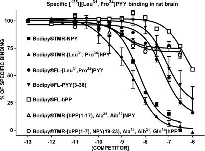
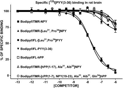
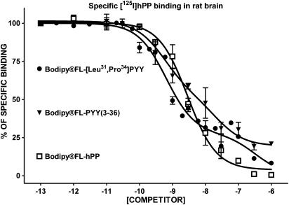
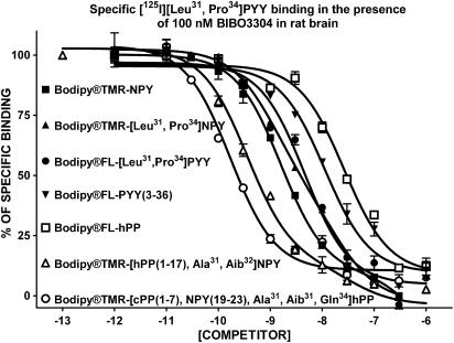
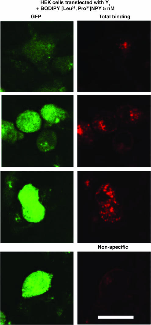
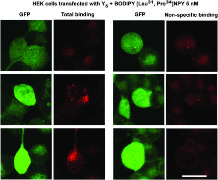
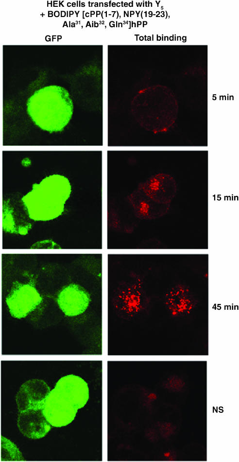
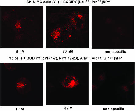
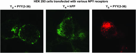
N-terminal labelled fluorescent BODIPY-NPY peptide analogues were tested in Y1, Y2, Y4 and Y5 receptor-binding assays performed in rat brain membrane preparations and HEK293 cells expressing the rat Y1, Y2, Y4 and Y5 receptors. BODIPY TMR/FL-[Leu31, Pro34]NPY/PYY were able to compete for specific [125][Leu31, Pro34]PYY-binding sites with an affinity similar to that observed for the native peptide at the Y1 (Ki=1-6 nM), Y2 (Ki>1000 nM), Y4 (Ki=10 nM) and Y5 (Ki=1-4 nM) receptor subtypes. BODIPY FL-PYY(3-36) was able to compete for specific Y2 (Ki=10 nM) and Y5 (Ki=30 nM) binding sites, but had almost no affinity in Y1 and Y4 assays. BODIPY FL-hPP was able to compete with high affinity (Ki; 1 and 15 nM) only in Y4 and Y5 receptor-binding assays. BODIPY TMR-[cPP(1-7), NPY(19-23), Ala31, Aib32, Gln34]hPP and BODIPY TMR-[hPP(1-17), Ala31, Aib32]NPY were potent competitors only on specific Y5-binding sites (Ki=0.1-0.6 nM). As expected, these fluorescent peptides inhibited forskolin-induced cAMP accumulation, demonstrating that they retained their agonist properties. When tested in confocal microscopy imaging, fluorescent Y1 and Y5 agonists internalized in a time-dependent manner in Y1 and Y5 transfected cells, respectively. These results demonstrate that BODIPY-conjugated NPY analogues retain their selectivity, affinity and agonist properties for the Y1, Y2, Y4 and Y5 receptor subtypes, respectively. Thus, they represent novel tools to study and visualize NPY receptors in living cells.


Figures

Figure 1
Figure 1
Competition-binding profiles of BODIPY®-labelled peptides against specific [125I][Leu31,Pro34]PYY-binding sites (Y1-, Y4- and Y5-like). Data are the mean±s.e.m. of two to five determinations, each performed in triplicate.
Figure 2
Figure 2
Competition-binding profiles of BODIPY®-labelled peptides against specific [125I]GR231118-binding sites (Y1-like). Data are the mean±s.e.m. of two to five determinations, each performed in triplicate.
Figure 3
Figure 3
Competition-binding profiles of BODIPY®-labelled peptides against specific [125I]PYY(3–36)-binding sites (Y2-like). Data are the mean±s.e.m. of two to five determinations, each performed in triplicate.
Figure 4
Figure 4
Competition-binding profiles of BODIPY®-labelled peptides against specific [125I]hPP-binding sites (Y4- and Y5-like). Data are the mean±s.e.m. of two to five determinations, each performed in triplicate.
Figure 5
Figure 5
Competition-binding profiles of BODIPY®-labelled peptides against specific [125I][Leu31,Pro34]PYY/BIBO3304-insensitive binding sites (Y4- and Y5-like). Data are the mean±s.e.m. of two to five determinations, each performed in triplicate.
Figure 6
Figure 6
Visualization of BODIPY®TMR-[Leu31, Pro34]NPY in HEK293 cells expressing the rat Y1 receptor incubated with 5 nM BODIPY®TMR-[Leu31, Pro34]NPY for 45 min at 37°C. Nonspecific binding represents signal obtained in the presence of 1 μM [Leu31, Pro34]NPY incubated under the same conditions with the fluorescent probe. The peptide is represented in red following HeNe laser (excitation 543 nm/emission 580 nm), and GFP-positive cells expressing low, moderate and high levels of GFP are represented in green following argon laser (excitation 488 nm/emission 510 nm). Scale bar 10 μm. All images were taken using the same setting.
Figure 7
Figure 7
Visualization of BODIPY®TMR-[Leu31, Pro34]NPY in HEK293 cells expressing the rat Y5 receptor incubated with 5 nM BODIPY®TMR-[Leu31, Pro34]NPY for 45 min at 37°C. Nonspecific binding represents signal obtained in the presence of 1 μM [Leu31, Pro34]NPY incubated under the same conditions with the fluorescent probe. The peptide is represented in red following HeNe laser (excitation 543 nm/emission 580 nm), and GFP-positive cells expressing low, moderate and high levels of GFP are represented in green following argon laser (excitation 488 nm/emission 510 nm). Scale bar 10 μm. All images were taken using the same setting.
Figure 8
Figure 8
Visualization of BODIPY®TMR-[cPP(1–7), NPY(19–23), Ala31, Aib32, Gln34]hPP in HEK293 cells expressing the rat Y5 receptor incubated for 5, 15 and 45 min at 37°C. Nonspecific binding represents signal obtained in the presence of 1 μM [cPP(1–7), NPY(19–23), Ala31, Aib32, Gln34]hPP incubated for 45 min with the fluorescent probe. The peptide is represented in red following HeNe laser (excitation 543 nm/emission 580 nm), and GFP-positive cells are represented in green following argon laser (excitation 488 nm/emission 510 nm). Scale bar 10 μm. All images were taken using the same setting.
Figure 9
Figure 9
Visualization of 5 and 20 nM BODIPY®TMR-[Leu31, Pro34]NPY in SK-N-MC cells and 1 and 5 nM BODIPY®TMR-[cPP(1–7), NPY(19–23), Ala31, Aib32, Gln34]hPP in HEK293 cells expressing the rat Y5 receptor. Cells were incubated with fluorescent probe for 45 min at 37°C. Nonspecific binding represents signal obtained in the presence of 1 μM native peptide incubated with the highest concentration of the fluorescent probe. Scale bar 10 μm. All images were taken using the same setting.
Figure 10
Figure 10
Visualization of BODIPY®FL-PYY(3–36), BODIPY®FL-hPP and BODIPY®TMR-PYY(3–36) in HEK293 cells expressing the rat Y2, Y4 or Y5 receptor cDNA, respectively. Y2-expressing cells were incubated with 20 nM BODIPY®FL-PYY(3–36) (excitation 488 nm/emission 510 nm), Y4 cells were incubated with 20 nM BODIPY®FL-hPP (excitation 488 nm/emission 510 nm), and Y5 cells with 5 nM BODIPY®TMR-PYY(3–36) (excitation 543 nm/emission 580 nm) for 45 min at 37°C. Nonspecific binding represents signal obtained in the presence of 1 μM native peptide incubated under the same conditions with the fluorescent probe. Scale bar 10 μm. All images were taken using the same setting.

Similar articles

Cited by

References

    1. ARTTAMANGKUL S., ALVAREZ-MAUBECIN V., THOMAS G., WILLIAMS J.T., GRANDY D.K. Binding and internalization of fluorescent opioid peptide conjugates in living cells. Mol. Pharmacol. 2000;58:1570–1580. - PubMed
    1. BARD J.A., WALKER M.W., BRANCHEK T.A., WEINSHANK R.L. Cloning and functional expression of a human Y4 subtype receptor for pancreatic polypeptide, neuropeptide Y, and peptide YY. J. Biol. Chem. 1995;270:26762–26765. - PubMed
    1. BECK-SICKINGER A.G., WIELAND H.A., WITTNEBEN H., WILLIM K.D., RUDOLF K., JUNG G. Complete L-alanine scan of neuropeptide Y reveals ligands binding to Y1 and Y2 receptors with distinguished conformations. Eur. J. Biochem. 1994;225:947–958. - PubMed
    1. BENNETT V.J., SIMMONS M.A. Analysis of fluorescently labeled substance P analogs: binding, imaging and receptor activation. BMC. Chem. Biol. 2001;1:1. - PMC - PubMed
    1. BERGLUND M.M., HIPSKIND P.A., GEHLERT D.R. Recent developments in our understanding of the physiological role of PP-fold peptide receptor subtypes. Exp. Biol. Med. (Maywood.). 2003a;228:217–244. - PubMed

Publication types

MeSH terms