. 2005 Dec;146(7):990-9.
doi: 10.1038/sj.bjp.0706410.

Activity of recombinant trypsin isoforms on human proteinase-activated receptors (PAR): mesotrypsin cannot activate epithelial PAR-1, -2, but weakly activates brain PAR-1

Affiliations

Activity of recombinant trypsin isoforms on human proteinase-activated receptors (PAR): mesotrypsin cannot activate epithelial PAR-1, -2, but weakly activates brain PAR-1

Zoryana Grishina et al. Br J Pharmacol. 2005 Dec.

Abstract

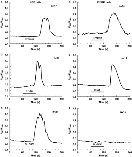
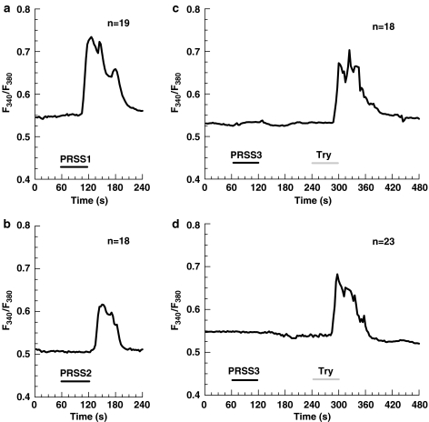
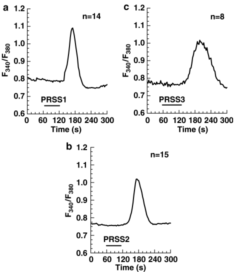
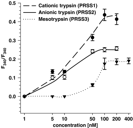
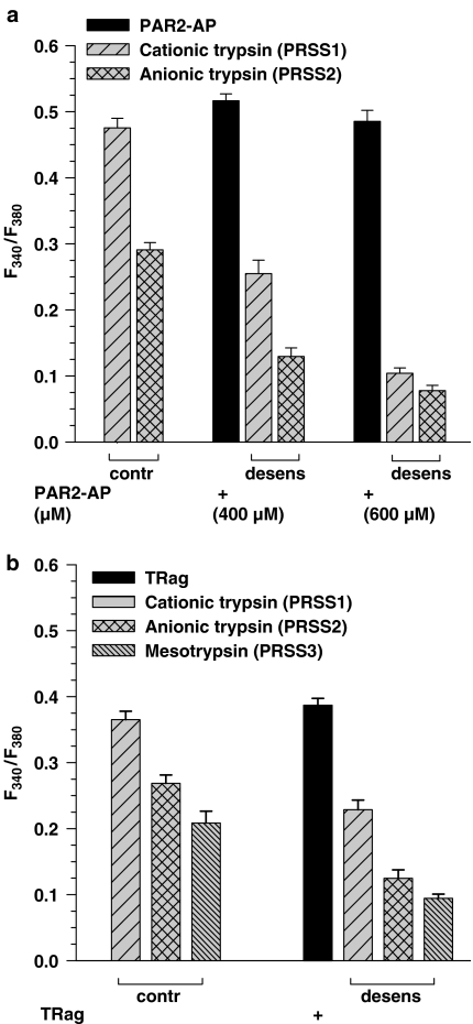
Trypsin-like serine proteinases trigger signal transduction pathways through proteolytic cleavage of proteinase-activated receptors (PARs) in many tissues. Three members, PAR-1, PAR-2 and PAR-4, are trypsin substrates, as trypsinolytic cleavage of the extracellular N terminus produces receptor activation. Here, the ability of the three human pancreatic trypsin isoforms (cationic trypsin, anionic trypsin and mesotrypsin (trypsin IV)) as recombinant proteins was tested on PARs. Using fura 2 [Ca(2+)](i) measurements, we analyzed three human epithelial cell lines, HBE (human bronchial epithelial), A549 (human pulmonary epithelial) and HEK (human embryonic kidney)-293 cells, which express functional PAR-1 and PAR-2. Human mesotrypsin failed to induce a PAR-mediated Ca(2+) response in human epithelial cells even at high concentrations. In addition, mesotrypsin did not affect the magnitude of PAR activation by subsequently added bovine trypsin. In HBE cells, which like A549 cells express high PAR-2 levels with negligible PAR-1 levels (<11%), half-maximal responses were seen for both cationic and anionic trypsins at about 5 nM. In the epithelial cells, mesotrypsin did not activate PAR-2 or PAR-1, whereas both anionic and cationic trypsins were comparable activators. We also investigated human astrocytoma 1321N1cells, which express PAR-1 and some PAR-3, but no PAR-2. High concentrations (>100 nM) of mesotrypsin produced a relatively weak Ca(2+) signal, apparently through PAR-1 activation. Half-maximal responses were observed at 60 nM mesotrypsin, and at 10-20 nM cationic and anionic trypsins. Using a desensitization assay with PAR-2-AP, we confirmed that both cationic and anionic trypsin isoforms cause [Ca(2+)](i) elevation in HBE cells mainly through PAR-2 activation. Desensitization of PAR-1 with thrombin receptor agonist peptide in 1321N1 cells demonstrated that all three recombinant trypsin isoforms act through PAR-1.Thus, the activity of human cationic and anionic trypsins on PARs was comparable to that of bovine pancreatic trypsin. Mesotrypsin (trypsin IV), in contrast to cationic and anionic trypsin, cannot activate or disable PARs in human epithelial cells, demonstrating that the receptors are no substrates for this isoenzyme. On the other hand, mesotrypsin activates PAR-1 in human astrocytoma cells. This might play a role in protection/degeneration or plasticity processes in the human brain.


Figures

Figure 1
Figure 1
Expression of proteinase-activated receptors in human cell lines: A549, HBE, HEK-293, 1321N1. PAR expression level in the cell lines determined by real-time RT–PCR. All mRNA measurements were normalized to the GAPDH mRNA level. The values given are means of triplicate measurement data±s.e. PAR expression levels are expressed relative to the PAR-1 mRNA expression in A549 cells, which was arbitrarily chosen as reference value of 1.
Figure 2
Figure 2
Ca2+ responses elicited in HBE (left) and 1321N1 (right) cells by: 10 nM and 50 nM commercial bovine trypsin (a and d, respectively), 1 μM PAR-1-AP TRAg (b and e), 100 μM PAR-2-AP SLIGKV (c and f). The cells were exposed to the agents as indicated by the respective bars. The changes of the intracellular calcium concentration ([Ca2+]i) in fura-2-AM loaded cells indicated by the change in the fluorescence ratio (F340 nm/F380 nm) were measured. The traces are the mean of the indicated number (n) of single cells measured in one experiment and are representative for at least three different experiments. The delay in the onset of the response is caused by the superfusion system.
Figure 3
Figure 3
Ca2+ responses in A549 cells produced by different isoforms of human recombinant trypsin: 10 nM cationic trypsin (PRSS1) (a), 10 nM anionic trypsin (PRSS2) (b), 10 nM mesotrypsin (PRSS3) followed by 10 nM bovine trypsin (c), and 400 nM mesotrypsin (PRSS3) followed by 10 nM bovine trypsin (d). The cells were exposed to the agents as indicated by the respective bars. The traces are the mean of the indicated number (n) of single cells measured in one experiment and are representative for at least three different experiments. The delay in the onset of the response is caused by the superfusion system.
Figure 4
Figure 4
Ca2+ responses in 1321N1 cells produced by different isoforms of human recombinant trypsin: 50 nM cationic trypsin (PRSS1) (a), 50 nM anionic trypsin (PRSS2) (b), 400 nM mesotrypsin (PRSS3) (c). The cells were exposed to the agents as indicated by the respective bars. The traces are the mean of the indicated number (n) of single cells measured in one experiment and are representative for at least three different experiments. The delay in the onset of the response is caused by the superfusion system.
Figure 5
Figure 5
Concentration–effect curves for recombinant human trypsin isoforms in human astrocytoma 1321N1 cells. The cells were stimulated for 1 min with varying concentrations of the proteases and the resulting change in the ratio of the fura 2 fluorescence was recorded. The amplitude values of the Ca2+ responses are given as means±s.e. from a minimum of 50 single cells measured in at least three different experiments (in some cases error bars are smaller in size than the symbols used).
Figure 6
Figure 6
Desensitization of Ca2+ responses in HBE (a) and 1321N1 (b) cells produced by the different isoforms of human recombinant trypsin after prestimulation of the cells with corresponding PAR-AP for 1 min. After 3 min washing, the cells were exposed for 1 min to a test concentration of the indicated trypsin isoforms. Control cells were not exposed to the peptide before the protease addition (left group of columns). HBE cells were treated with 400 or 600 μM PAR-2-AP, followed by the application of 10 nM of cationic or anionic trypsins (a). 1321N1 cells were treated with 10 μM TRag, followed by the application of 50 nM cationic or anionic trypsins or 400 nM mesotrypsin (b). The amplitude values of the Ca2+ responses are given as means±s.e.m. from a minimum of 50 single cells measured in at least three different experiments with the experimental protocol presented.

Similar articles

Cited by

References

    1. BLACKHART B.D., EMILSSON K., NGUYEN D., TENG W., MARTELLI A.J., NYSTEDT S., SUNDELIN J., SCARBOROUGH R.M. Ligand cross-reactivity within the protease-activated receptor family. J. Biol. Chem. 1996;271:16466–16471. - PubMed
    1. COMPTON S.J., CAIRNS J.A., PALMER K.J., AL-ANI B., HOLLENBERG M.D., WALLS A.F. A polymorphic protease-activated receptor 2 (PAR2) displaying reduced sensitivity to trypsin and differential responses to PAR agonists. J. Biol. Chem. 2000;275:39207–39212. - PubMed
    1. COMPTON S.J., RENAUX B., WIJESURIYA S.J., HOLLENBERG M.D. Glycosylation and the activation of proteinase-activated receptor 2 (PAR(2)) by human mast cell tryptase. Br. J. Pharmacol. 2001;134:705–718. - PMC - PubMed
    1. COTTRELL G.S., AMADESI S., GRADY E.F., BUNNETT N.W. Trypsin IV: A novel agonist of protease-activated receptors 2 and 4. J. Biol. Chem. 2004;279:13532–13539. - PubMed
    1. DERY O., CORVERA C.U., STEINHOFF M., BUNNETT N.W. Proteinase-activated receptors: novel mechanisms of signaling by serine proteases. Am. J. Physiol. 1998;274:C1429–C1452. - PubMed

Publication types