

Oxaliplatin induces hyperexcitability at motor and autonomic neuromuscular junctions through effects on voltage-gated sodium channels
- PMID: 16231011
- PMCID: PMC1751225
- DOI: 10.1038/sj.bjp.0706407
Oxaliplatin induces hyperexcitability at motor and autonomic neuromuscular junctions through effects on voltage-gated sodium channels
Abstract
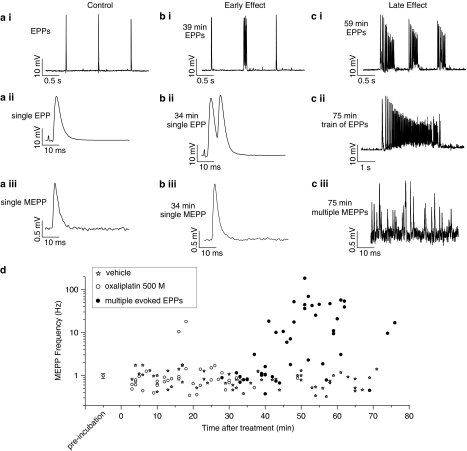
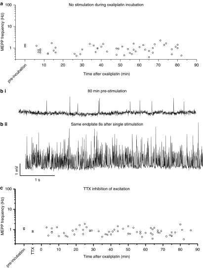
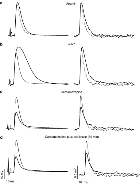
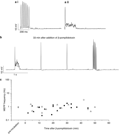
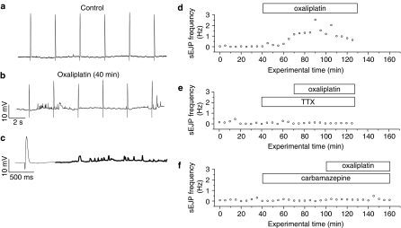
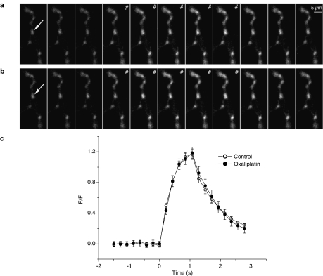
Oxaliplatin, an effective cytotoxic treatment in combination with 5-fluorouracil for colorectal cancer, is associated with sensory, motor and autonomic neurotoxicity. Motor symptoms include hyperexcitability while autonomic effects include urinary retention, but the cause of these side-effects is unknown. We examined the effects on motor nerve function in the mouse hemidiaphragm and on the autonomic system in the vas deferens. In the mouse diaphragm, oxaliplatin (0.5 mM) induced multiple endplate potentials (EPPs) following a single stimulus, and was associated with an increase in spontaneous miniature EPP frequency. In the vas deferens, spontaneous excitatory junction potential frequency was increased after 30 min exposure to oxaliplatin; no changes in resting Ca(2+) concentration in nerve terminal varicosities were observed, and recovery after stimuli trains was unaffected. In both tissues, an oxaliplatin-induced increase in spontaneous activity was prevented by the voltage-gated Na(+) channel blocker tetrodotoxin (TTX). Carbamazepine (0.3 mM) also prevented multiple EPPs and the increase in spontaneous activity in both tissues. In diaphragm, beta-pompilidotoxin (100 microM), which slows Na(+) channel inactivation, induced multiple EPPs similar to oxaliplatin's effect. By contrast, blockers of K(+) channels (4-aminopyridine and apamin) did not replicate oxaliplatin-induced hyperexcitability in the diaphragm. The prevention of hyperexcitability by TTX blockade implies that oxaliplatin acts on nerve conduction rather than by effecting repolarisation. The similarity between beta-pompilidotoxin and oxaliplatin suggests that alteration of voltage-gated Na(+) channel kinetics is likely to underlie the acute neurotoxic actions of oxaliplatin.
Figures
Similar articles
-
The chemotherapeutic oxaliplatin alters voltage-gated Na(+) channel kinetics on rat sensory neurons.Eur J Pharmacol. 2000 Oct 6;406(1):25-32. doi: 10.1016/s0014-2999(00)00667-1. Eur J Pharmacol. 2000. PMID: 11011028
-
A possible explanation for a neurotoxic effect of the anticancer agent oxaliplatin on neuronal voltage-gated sodium channels.J Neurophysiol. 2001 May;85(5):2293-7. doi: 10.1152/jn.2001.85.5.2293. J Neurophysiol. 2001. PMID: 11353042
-
A new conotoxin isolated from Conus consors venom acting selectively on axons and motor nerve terminals through a Na+-dependent mechanism.Eur J Neurosci. 1999 Sep;11(9):3134-42. doi: 10.1046/j.1460-9568.1999.00732.x. Eur J Neurosci. 1999. PMID: 10510177
-
Clinical aspects and molecular basis of oxaliplatin neurotoxicity: current management and development of preventive measures.Semin Oncol. 2002 Oct;29(5 Suppl 15):21-33. doi: 10.1053/sonc.2002.35525. Semin Oncol. 2002. PMID: 12422305 Review.
-
[Painful hyperexcitability syndrome with oxaliplatin containing chemotherapy. Clinical features, pathophysiology and therapeutic options].Schmerz. 2008 Feb;22(1):16-23. doi: 10.1007/s00482-007-0552-5. Schmerz. 2008. PMID: 17578604 Review. German.
Cited by
-
Calcium and magnesium prophylaxis for oxaliplatin-related neurotoxicity: is it a trade-off between drug efficacy and toxicity?Oncologist. 2011;16(12):1780-3. doi: 10.1634/theoncologist.2011-0157. Epub 2011 Nov 29. Oncologist. 2011. PMID: 22128115 Free PMC article. Review.
-
Mechanisms involved in the development of chemotherapy-induced neuropathy.Pain Manag. 2015;5(4):285-96. doi: 10.2217/pmt.15.19. Epub 2015 Jun 19. Pain Manag. 2015. PMID: 26087973 Free PMC article. Review.
-
Ion channels and neuronal hyperexcitability in chemotherapy-induced peripheral neuropathy; cause and effect?Mol Pain. 2017 Jan-Dec;13:1744806917714693. doi: 10.1177/1744806917714693. Mol Pain. 2017. PMID: 28580836 Free PMC article.
-
Platinum-induced neurotoxicity: A review of possible mechanisms.World J Clin Oncol. 2017 Aug 10;8(4):329-335. doi: 10.5306/wjco.v8.i4.329. World J Clin Oncol. 2017. PMID: 28848699 Free PMC article. Review.
-
Targeting Mitochondria and Oxidative Stress in Cancer- and Chemotherapy-Induced Muscle Wasting.Antioxid Redox Signal. 2023 Feb;38(4-6):352-370. doi: 10.1089/ars.2022.0149. Epub 2022 Dec 29. Antioxid Redox Signal. 2023. PMID: 36310444 Free PMC article. Review.
References
-
- ADELSBERGER H., QUASTHOFF S., GROSSKREUTZ J., LEPIER A., ECKEL F., LERSCH C. The chemotherapeutic oxaliplatin alters voltage-gated Na+ channel kinetics on rat sensory neurons. Eur. J. Pharmacol. 2000;406:25–32. - PubMed
-
- ANDRE T., BONI C., MOUNEDJI-BOUDIAF L., NAVARRO M., TABERNERO J., HICKISH T., TOPHAM C., ZANINELLI M., CLINGAN P., BRIDGEWATER J., TABAH-FISCH I., DE GRAMONT A. Oxaliplatin, fluorouracil, and leucovorin as adjuvant treatment for colon cancer. N. Engl. J. Med. 2004;350:2343–2351. - PubMed
-
- BRAIN K.L., TROUT S.J., JACKSON V.M., DASS N., CUNNANE T.C. Nicotine induces calcium spikes in single nerve terminal varicosities: a role for intracellular calcium stores. Neuroscience. 2001;106:395–403. - PubMed
Publication types
MeSH terms
Substances
Grants and funding
LinkOut - more resources
Full Text Sources
Other Literature Sources
Miscellaneous
