. 2006 Feb 15;394(Pt 1):43-50.
doi: 10.1042/BJ20051419.

Intracellular composition of fatty acid affects the processing and function of tyrosinase through the ubiquitin-proteasome pathway

Affiliations

Intracellular composition of fatty acid affects the processing and function of tyrosinase through the ubiquitin-proteasome pathway

Hideya Ando et al. Biochem J. .

Abstract

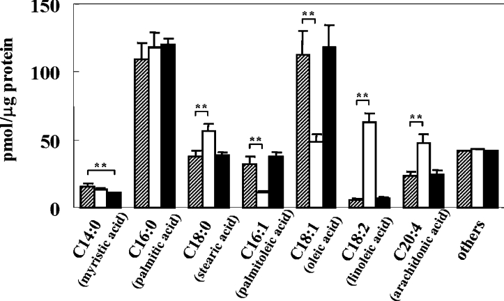
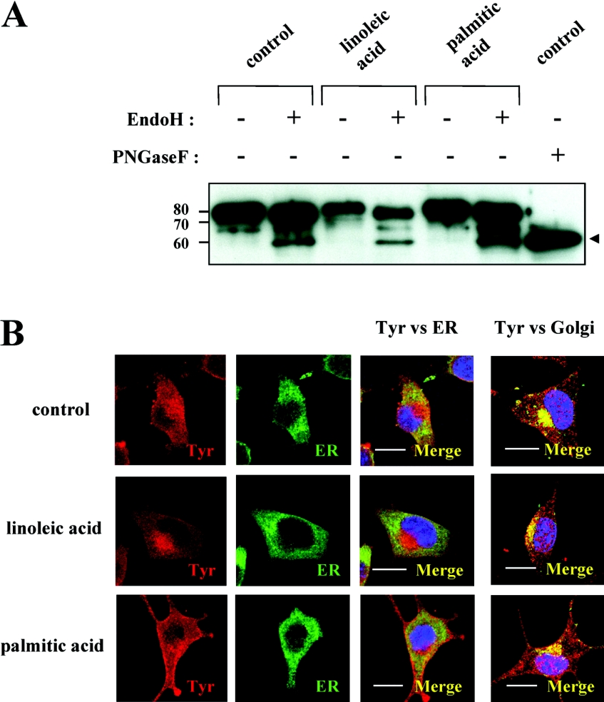
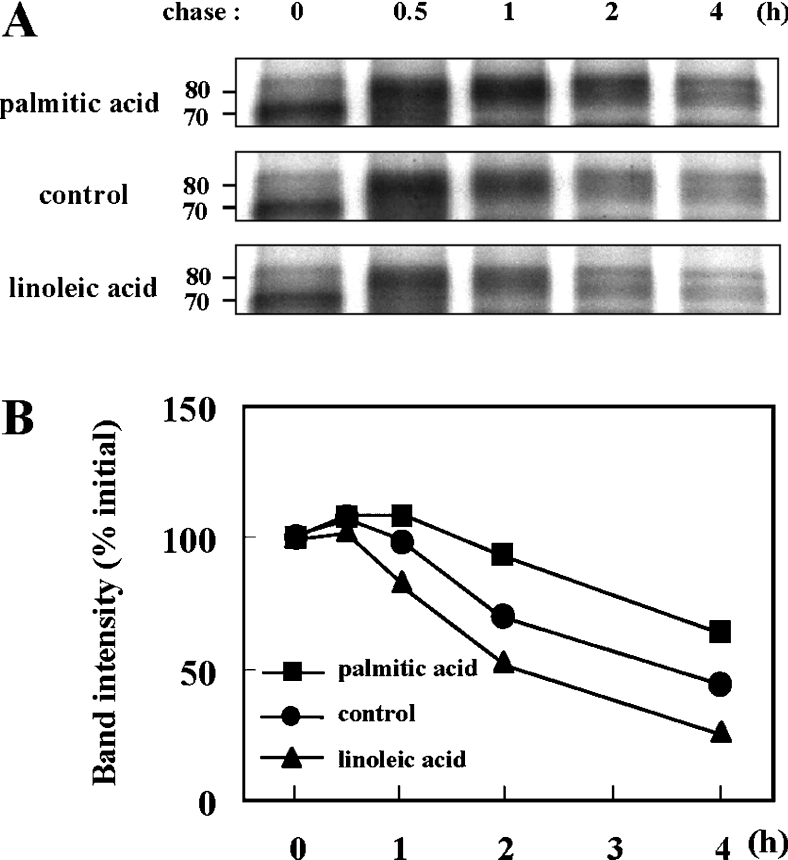
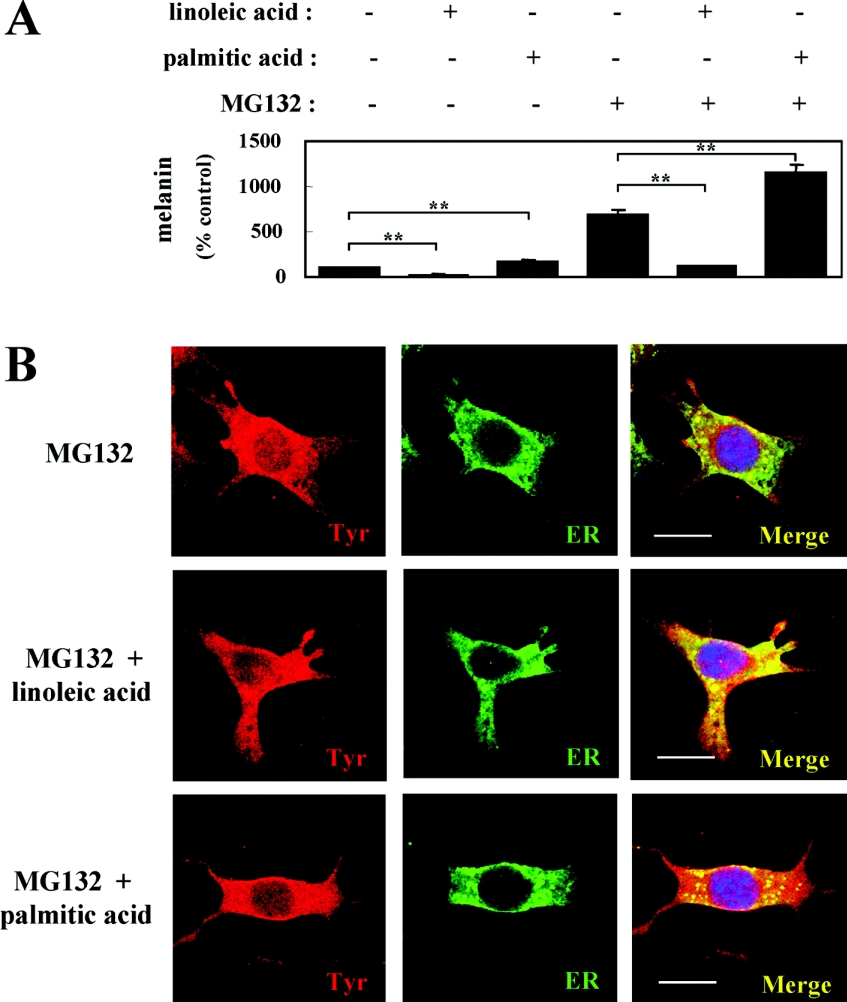
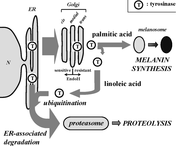
Proteasomes are multicatalytic proteinase complexes within cells that selectively degrade ubiquitinated proteins. We have recently demonstrated that fatty acids, major components of cell membranes, are able to regulate the proteasomal degradation of tyrosinase, a critical enzyme required for melanin biosynthesis, in contrasting manners by relative increases or decreases in the ubiquitinated tyrosinase. In the present study, we show that altering the intracellular composition of fatty acids affects the post-Golgi degradation of tyrosinase. Incubation with linoleic acid (C18:2) dramatically changed the fatty acid composition of cultured B16 melanoma cells, i.e. the remarkable increase in polyunsaturated fatty acids such as linoleic acid and arachidonic acid (C20:4) was compensated by the decrease in monounsaturated fatty acids such as oleic acid (C18:1) and palmitoleic acid (C16:1), with little effect on the proportion of saturated to unsaturated fatty acid. When the composition of intracellular fatty acids was altered, tyrosinase was rapidly processed to the Golgi apparatus from the ER (endoplasmic reticulum) and the degradation of tyrosinase was increased after its maturation in the Golgi. Retention of tyrosinase in the ER was observed when cells were treated with linoleic acid in the presence of proteasome inhibitors, explaining why melanin synthesis was decreased in cells treated with linoleic acid and a proteasome inhibitor despite the abrogation of tyrosinase degradation. These results suggest that the intracellular composition of fatty acid affects the processing and function of tyrosinase in connection with the ubiquitin-proteasome pathway and suggest that this might be a common physiological approach to regulate protein degradation.


Figures

Figure 1
Figure 1. Incubation of linoleic acid dramatically alters the intracellular composition of fatty acids
Bar graphs of fatty acid composition of B16 melanoma cells after incubation with 25 μM linoleic acid (open bar) or palmitic acid (closed bar) for 72 h, as compared with the control (shaded bar). Results shown are mean±S.D. for triplicate determinations. Student's t test was used for statistical analysis of the data (**P<0.01 versus the control).
Figure 2
Figure 2. Tyrosinase is rapidly processed to the Golgi with little change in sensitivity to glycosidase digestion
(A) Western blotting of tyrosinase with (+) or without (−) glycosidase digestion with endo H or PNGase F in whole cell lysates (1 μg of protein) of B16 melanoma cells pretreated with linoleic acid (25 μM) or palmitic acid (25 μM) for 72 h. Arrowhead indicates the position of endo H-sensitive and PNGase F-digested band. Numbers on the left indicate masses of protein in kDa. (B) Immunofluorescence confocal microscopy showing the intracellular distribution of tyrosinase, ER and Golgi after incubation with fatty acids. B16 mouse melanoma cells were treated with linoleic acid (25 μM; middle row panels), palmitic acid (25 μM; lower row panels) or DMSO only (control; upper row panels) for 72 h. Cells were stained with antibodies for tyrosinase (αPEP7 at 1:20), ER (Bip/GRP78 at 1:10) or Golgi (Vti1b at 1:10). Reactivity was classified into three categories, according to whether they showed red (tyrosinase), green (ER) or yellow fluorescence. The left two column panels show tyrosinase (Tyr) and ER respectively, while the right two column panels represent the merged images indicating co-localization of tyrosinase with the ER and Golgi respectively as yellow fluorescence. Three independent experiments were performed with similar results and panels showing only single representative cells are shown. Scale bar, 10 μm.
Figure 3
Figure 3. Fatty acids regulate the degradation of the glycosylated form of mature tyrosinase
(A) Metabolic labelling and immunoprecipitation analysis of tyrosinase after pretreatment with linoleic acid (25 μM) or palmitic acid (25 μM) for 72 h, showing the processing and degradation of tyrosinase in B16 melanoma cells. Cells were pulsed for 30 min and then chased for the indicated times. Numbers on the left indicate masses of protein in kDa. Three independent experiments were performed and representative blots are shown. (B) Line graphs of band intensities of immature (0 h, 70 kDa) and glycosylated (0.5–4 h, 80 kDa) tyrosinase pretreated with linoleic acid (▲) or palmitic acid (■) or untreated controls (●). The intensities of the 70 and 80 kDa bands were measured individually. The initial intensity of each band (0 h) is adjusted to 100% and the relative intensities are recalculated to determine tyrosinase degradation. Shown are representative results from three independent experiments.
Figure 4
Figure 4. ER retention of tyrosinase is elicited by linoleic acid incubation when proteasome activity is inhibited
(A) Melanin measurements of B16 melanoma cells after incubation with (+) or without (−) fatty acids, in the presence (+) or absence (−) of MG132, for 72 h. Results shown are mean±S.D. for triplicate determinations. Student's t test was used for statistical analysis of the data (**P<0.01 versus untreated control or MG132-treated control). (B) Immunofluorescence confocal microscopy showing the intracellular distribution of tyrosinase and the ER after incubation with fatty acids in the presence of MG132. B16 melanoma cells were treated with linoleic acid (25 μM; middle row panels), palmitic acid (25 μM; lower row panels) or DMSO only (control; upper row panels) for 72 h in the presence of MG132 (120 nM) for the last 24 h of treatment before fixation. The merged images indicate co-localization of tyrosinase and the ER as yellow fluorescence. Three independent experiments were performed with similar results and panels showing only single representative cells are shown. Scale bar, 10 μm.
Figure 5
Figure 5. Scheme depicting the trafficking of tyrosinase after incubation with linoleic acid or palmitic acid
After rapid processing of tyrosinase from the ER to the Golgi, linoleic acid enhances the ubiquitination of mature tyrosinase and the ubiquitinated tyrosinase could be integrated into the ERAD. In contrast, palmitic acid diminishes the proteasomal degradation of tyrosinase, leading to melanin formation in melanosomes.

Similar articles

Cited by

References

    1. Tsujishita Y., Asaoka Y., Nishizuka Y. Regulation of phospholipase A2 in human leukemia cell lines: its implication for intracellular signaling. Proc. Natl. Acad. Sci. U.S.A. 1994;91:6274–6278. - PMC - PubMed
    1. Chawla A., Repa J. J., Evans R. M., Mangelsdorf D. J. Nuclear receptors and lipid physiology: opening the X-files. Science. 2001;294:1866–1870. - PubMed
    1. Clarke S. D. The multi-dimensional regulation of gene expression by fatty acids: polyunsaturated fats as nutrient sensors. Curr. Opin. Lipidol. 2004;15:13–18. - PubMed
    1. Tahin Q. S., Blum M., Carafoli E. The fatty acid composition of subcellular membranes of rat liver, heart, and brain: diet-induced modifications. Eur. J. Biochem. 1981;121:5–13. - PubMed
    1. Awad A. B., Chattopadhyay J. P. Effect of dietary saturated fatty acids on intracellular free fatty acids and kinetic properties of hormone-sensitive lipase of rat adipocytes. J. Nutr. 1986;116:1095–1100. - PubMed

Publication types