

The costimulatory molecules CD80, CD86 and OX40L are up-regulated in Aspergillus fumigatus sensitized mice
- PMID: 16232210
- PMCID: PMC1809515
- DOI: 10.1111/j.1365-2249.2005.02905.x
The costimulatory molecules CD80, CD86 and OX40L are up-regulated in Aspergillus fumigatus sensitized mice
Abstract
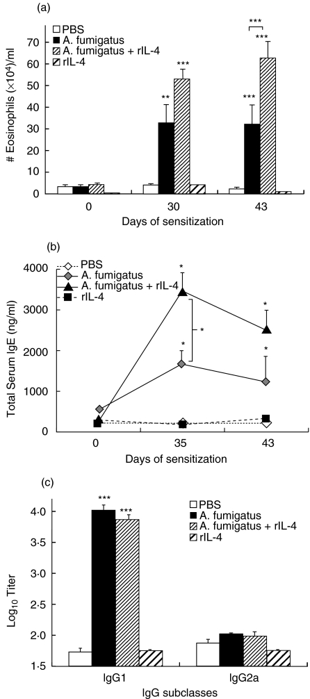
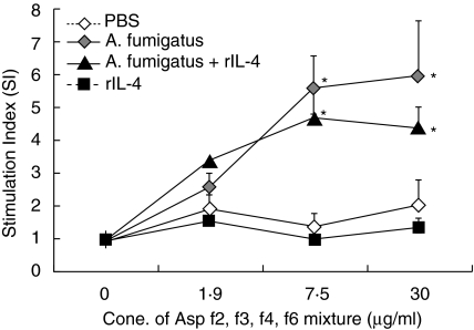
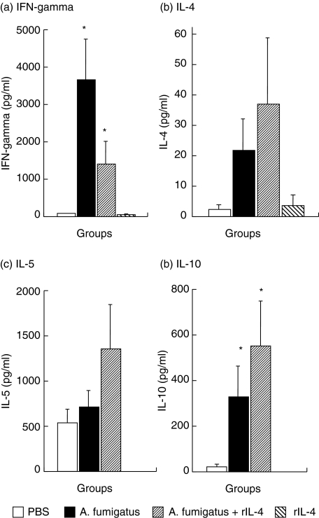
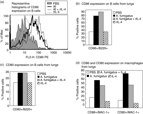
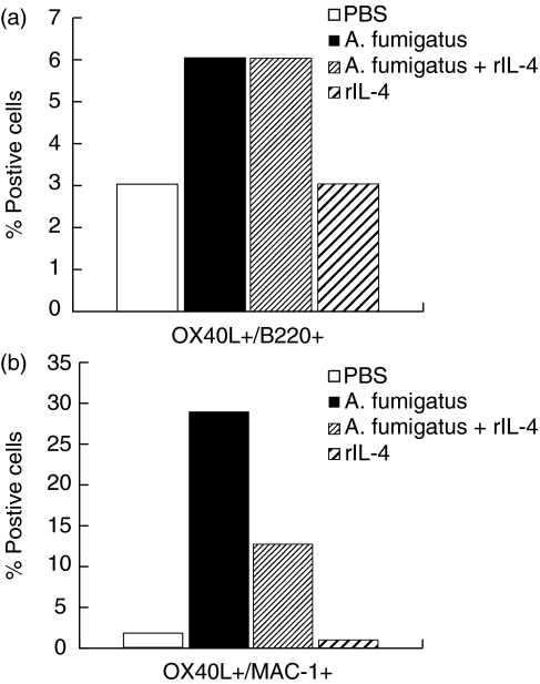
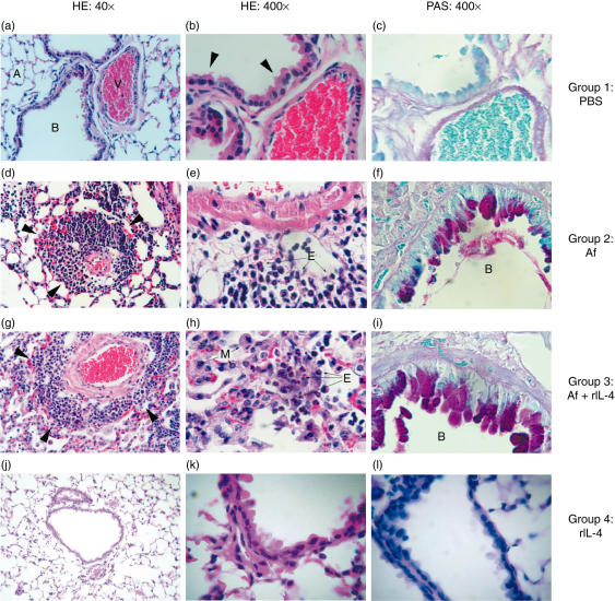
Aspergillus fumigatus (Af) is a fungus associated with allergic bronchopulmonary aspergillosis (ABPA) and other allergic diseases. Immune responses in these diseases are due to T and B cell responses. T cell activation requires both Af-specific engagement of the T-cell-receptor as well as interaction of antigen independent costimulatory molecules including CD28-CD80/CD86 and OX40-OX40L interactions. Since these molecules and their interactions have been suggested to have a potential involvement in the pathogenesis of ABPA, we have investigated their role in a model of experimental allergic aspergillosis. BALB/c mice were primed and sensitized with Af allergens, with or without exogenous IL-4. Results showed up-regulation of both CD86 and CD80 molecules on lung B cells from Af-sensitized mice (79% CD86+ and 24% CD80+) and Af/rIL-4-treated mice (90% CD86+ and 24% CD80+) compared to normal controls (36% and 17%, respectively). Lung macrophages in Af-sensitized mice treated or not with IL-4 showed enhanced expression of these molecules. OX40L expression was also up-regulated on lung B cells and macrophages from both Af-sensitized and Af/rIL-4 exposed mice as compared to normal controls. All Af-sensitized animals showed peripheral blood eosinophilia, enhanced total serum IgE and allergen-specific IgG1 antibodies and characteristic lung inflammation. The up-regulation of CD80, CD86 and OX40L molecules on lung B cells and macrophages from Af-allergen exposed mice suggests a major role for these molecules in the amplification and persistence of immunological and inflammatory responses in ABPA.
Figures
Similar articles
-
Molecular-based allergy diagnosis of allergic bronchopulmonary aspergillosis in Aspergillus fumigatus-sensitized Japanese patients.Clin Exp Allergy. 2015 Dec;45(12):1790-800. doi: 10.1111/cea.12590. Clin Exp Allergy. 2015. PMID: 26118958
-
CD28 interactions with either CD80 or CD86 are sufficient to induce allergic airway inflammation in mice.Am J Respir Cell Mol Biol. 1999 Oct;21(4):498-509. doi: 10.1165/ajrcmb.21.4.3714. Am J Respir Cell Mol Biol. 1999. PMID: 10502560
-
Modulation of airway inflammation by immunostimulatory CpG oligodeoxynucleotides in a murine model of allergic aspergillosis.Infect Immun. 2004 Oct;72(10):6087-94. doi: 10.1128/IAI.72.10.6087-6094.2004. Infect Immun. 2004. PMID: 15385513 Free PMC article.
-
Immunology of allergic bronchopulmonary aspergillosis.Indian J Chest Dis Allied Sci. 2000 Oct-Dec;42(4):225-37. Indian J Chest Dis Allied Sci. 2000. PMID: 15597669 Review.
-
Molecular characterization of aspergillus fumigatus allergens.Indian J Chest Dis Allied Sci. 2000 Oct-Dec;42(4):239-48. Indian J Chest Dis Allied Sci. 2000. PMID: 15597670 Review.
Cited by
-
Reverse Signaling of Tumor Necrosis Factor Superfamily Proteins in Macrophages and Microglia: Superfamily Portrait in the Neuroimmune Interface.Front Immunol. 2019 Feb 19;10:262. doi: 10.3389/fimmu.2019.00262. eCollection 2019. Front Immunol. 2019. PMID: 30838001 Free PMC article. Review.
-
Altered expression of TNFSF4 and TRAF2 mRNAs in peripheral blood mononuclear cells in patients with systemic lupus erythematosus: association with atherosclerotic symptoms and lupus nephritis.Inflamm Res. 2012 Dec;61(12):1347-54. doi: 10.1007/s00011-012-0535-6. Epub 2012 Jul 31. Inflamm Res. 2012. PMID: 22847298
-
Immune response among patients exposed to molds.Int J Mol Sci. 2009 Dec 21;10(12):5471-84. doi: 10.3390/ijms10125471. Int J Mol Sci. 2009. PMID: 20054481 Free PMC article.
-
Pan-cancer analysis shows that TNFSF4 is a potential prognostic and immunotherapeutic biomarker for multiple cancer types including liver cancer.BMC Cancer. 2025 Jan 17;25(1):100. doi: 10.1186/s12885-025-13479-4. BMC Cancer. 2025. PMID: 39825223 Free PMC article.
-
Antagonism of airway tolerance by endotoxin/lipopolysaccharide through promoting OX40L and suppressing antigen-specific Foxp3+ T regulatory cells.J Immunol. 2008 Dec 15;181(12):8650-9. doi: 10.4049/jimmunol.181.12.8650. J Immunol. 2008. PMID: 19050285 Free PMC article.
References
-
- Greenberger PA. Middletonís Allergy Principles and Practice. In: Adkinson NF Jr, Yunginger JW, Busse WW, Bochner BS, Holgate ST, Simons FER, editors. St Louis: Mosby; 2003. pp. 1353–71.
-
- Knutsen AP, Chauhan B, Slavin RG. Cell-mediated immunity in allergic bronchopulmonary aspergillosis. In: Kurup VP, Apter AJ, editors. Immunology and Allergy Clinics of North America. Philadelphia: W.B. Saunders; 1998. pp. 575–99.
-
- Kurup VP, Madan T, Sarma UP. Allergic bronchopulmonary aspergillosis. Recent concepts and considerations. In: Domer JE, Kobayashi GS, editors. The Mycota XII. Human Fungal Pathogens. Berlin: Springer-Verlag; 2004. pp. 225–41.
-
- Abbas AK. The control of T cell activation vs. tolerance. Autoimmun Rev. 2003;2:115–8. - PubMed
-
- Greenwald RJ, Freeman GJ, Sharpe AH. The B7 family revisited. Annu Rev Immunol. 2005;23:515–48. - PubMed
Publication types
MeSH terms
Substances
LinkOut - more resources
Full Text Sources
