. 2005 Oct 18:2:62.
doi: 10.1186/1742-4690-2-62.

Contribution of the C-terminal tri-lysine regions of human immunodeficiency virus type 1 integrase for efficient reverse transcription and viral DNA nuclear import

Affiliations

Contribution of the C-terminal tri-lysine regions of human immunodeficiency virus type 1 integrase for efficient reverse transcription and viral DNA nuclear import

Zhujun Ao et al. Retrovirology. .

Abstract

Background: In addition to mediating the integration process, HIV-1 integrase (IN) has also been implicated in different steps during viral life cycle including reverse transcription and viral DNA nuclear import. Although the karyophilic property of HIV-1 IN has been well demonstrated using a variety of experimental approaches, the definition of domain(s) and/or motif(s) within the protein that mediate viral DNA nuclear import and its mechanism are still disputed and controversial. In this study, we performed mutagenic analyses to investigate the contribution of different regions in the C-terminal domain of HIV-1 IN to protein nuclear localization as well as their effects on virus infection.

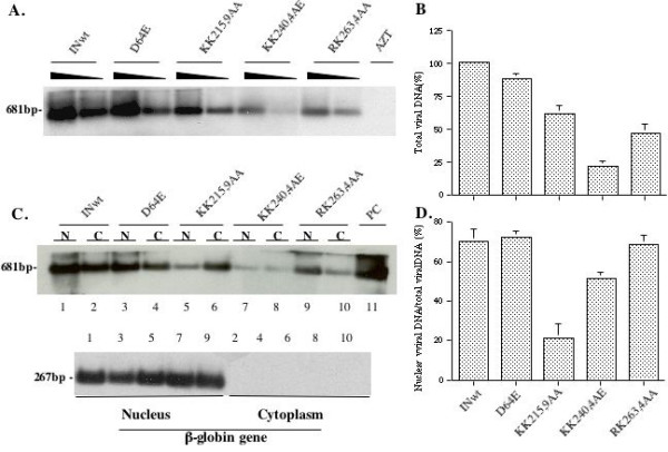
Results: Our analysis showed that replacing lysine residues in two highly conserved tri-lysine regions, which are located within previously described Region C (235WKGPAKLLWKGEGAVV) and sequence Q (211KELQKQITK) in the C-terminal domain of HIV-1 IN, impaired protein nuclear accumulation, while mutations for RK263,4 had no significant effect. Analysis of their effects on viral infection in a VSV-G pseudotyped RT/IN trans-complemented HIV-1 single cycle replication system revealed that all three C-terminal mutant viruses (KK215,9AA, KK240,4AE and RK263,4AA) exhibited more severe defect of induction of beta-Gal positive cells and luciferase activity than an IN class 1 mutant D64E in HeLa-CD4-CCR5-beta-Gal cells, and in dividing as well as non-dividing C8166 T cells, suggesting that some viral defects are occurring prior to viral integration. Furthermore, by analyzing viral DNA synthesis and the nucleus-associated viral DNA level, the results clearly showed that, although all three C-terminal mutants inhibited viral reverse transcription to different extents, the KK240,4AE mutant exhibited most profound effect on this step, whereas KK215,9AA significantly impaired viral DNA nuclear import. In addition, our analysis could not detect viral DNA integration in each C-terminal mutant infection, even though they displayed various low levels of nucleus-associated viral DNA, suggesting that these C-terminal mutants also impaired viral DNA integration ability.

Conclusion: All of these results indicate that, in addition to being involved in HIV-1 reverse transcription and integration, the C-terminal tri-lysine regions of IN also contribute to efficient viral DNA nuclear import during the early stage of HIV-1 replication.


Figures

Figure 1
Figure 1
Subcellular localization of the wild-type and truncated HIV integrase fused with YFP. A) Schematic structure of HIV-1 integrase-YFP fusion proteins. Full-length (1–288aa) HIV-1 integrase, the N-terminus-truncated mutant (51–228aa) or the C-terminus-truncated mutant (1–212aa) was fused in frame at the N-terminus of YFP protein. The cDNA encoding for each IN-YFP fusion protein was inserted in a SVCMV expression plasmid. B) Expression of different IN-YFP fusion proteins in 293T cells. 293T cells were transfected with each IN-YFP expressor and at 48 hours of transfection, cells were lysed, immunoprecipitated with anti-HIV serum and resolved by electrophoresis through a 12.5% SDS-PAGE followed by Western blot with rabbit anti-GFP antibody. The molecular weight markers are indicated at the left side of the gel. C) Intracellular localization of different IN-YFP fusion proteins. HeLa cells were transfected with each HIV-1 IN-YFP fusion protein expressor and at 48 hours of transfection, cells were fixed and subjected to indirect immunofluorescence using rabbit anti-GFP and then incubated with FITC-conjugated anti-rabbit antibodies. The localization of each fusion protein was viewed by Fluorescence microscopy with a 50× oil immersion objective. Upper panel is fluorescence images and bottom panel is DAPI nucleus staining.
Figure 2
Figure 2
Effect of different IN C-terminal substitution mutants on IN-YFP intracellular localization. A) Diagram of HIV-1 IN domain structure and introduced mutations at the C-terminal domain of the protein. The position of lysines in two tri-lysine regions and introduced mutations are shown at the bottom of sequence. B) The expression of the wild-type and mutant IN-YFP fusion proteins were detected in transfected 293T cells by using immunoprecipitation with anti-HIV serum and Western blot with rabbit anti-GFP antibody, as described in figure 1. The molecular weight markers are indicated at the left side of the gel. C) Intracellular localization of different HIV-1 IN mutant-YFP fusion proteins in HeLa cells were analyzed by fluorescence microscopy with a 50× oil immersion objective. The nucleus of HeLa cells was simultaneously visualized by DAPI staining (lower panel).
Figure 3
Figure 3
Production of different single-cycle replicating viruses and their infection in HeLa-CD4-CCR5-β-Gal cells. A). To evaluate the trans-incorporation of RT and IN in VSV-G pseudotyped viral particles, viruses released from 293T cells transfected with NLlucΔBglΔRI provirus alone (lane 6) or cotransfected with different Vpr-RT-IN expressors and a VSV-G expressor (lane 1 to 5) were lysed, immunoprecipitated with anti-HIV serum. Then, immunoprecipitates were run in 12% SDS-PAGE and analyzed by Western blot with rabbit anti-IN antibody (middle panel) or anti-RT and anti-p24 monoclonal antibody (upper and lower panel). B) The infectivity of trans-complemented viruses produced in 293 T cells was evaluated by MAGI assay. HeLa-CD4-CCR5-LTR-β-Gal cells were infected with equal amounts (at 10 cpm/cell) of different IN mutant viruses and after 48 hours of infection, numbers of β-Gal positive cells (infected cell) were monitored by X-gal staining. Error bars represent variation between duplicate samples and the data is representative of results obtained in three independent experiments.
Figure 4
Figure 4
Effect of IN mutants on viral infection in dividing and nondividing C8166 T cells. To test the effect of different IN mutants on HIV-1 infection in CD4+ T cells, dividing (panel A) and non-dividing (aphidicolin-treated, panel B) C8166 T cells were infected with equal amount of VSV-G pseudotyped IN mutant viruses (at 5 cpm/cell). For evaluation of the effect of different IN mutants on HIV-1 envelope-mediated infection in CD4+ T cells, dividing C8166 T cells were infected with equal amount of HIV-1 envelope competent IN mutant viruses (at 10 cpm/cell) (panel C). After 48 hours of infection, HIV-1 DNA-mediated luciferase induction was monitored by luciferase assay. Briefly, the same amount (106 cells) of cells was lysed in 50 ul of luciferase lysis buffer and then, 10 μl of cell lysate was subjected to the luciferase assay. Error bars represent variation between duplicate samples and the data is representative of results obtained in three independent experiments.
Figure 5
Figure 5
Effects of different IN mutants on HIV-1 reverse transcription and DNA nuclear import. Dividing C8166 T cells were infected with equal amounts of different HIV-1 IN mutant viruses. A) At 12 hours post-infection, 1 × 10⁶ cells were lysed and the total viral DNA was detected by PCR using HIV-1 LTR-Gag primers and Southern blot. B) Levels of HIV-1 late reverse transcription products detected in panel A were quantified by laser densitometry and viral DNA level of the wt virus was arbitrarily set as 100%. Means and standard deviations from two independent experiments are presented. C) At 24 hours post-infection, 2 × 10⁶ cells were fractionated into cytoplasmic and nuclear fractions as described in Materials and Methods. The amount of viral DNA in cytoplasmic and nuclear fractions were analyzed by PCR using HIV-1 LTR-Gag primers and Southern blot (upper panel, N. nuclear fraction; C. cytoplasmic fraction). Purity and DNA content of each subcellular fraction were monitored by PCR detection of human globin DNA and visualized by specific Southern blot (lower panel). D). The percentage of nucleus-associated viral DNA relative to the total amount of viral DNA for each mutant was also quantified by laser densitometry. Means and standard deviations from two independent experiments are shown.
Figure 6
Figure 6
Effect of IN mutants on HIV-1 proviral DNA integration. Dividing C8166 T cells were infected with equal amounts of different HIV-1 IN mutant viruses. At 24 hours post-infection, 1 × 10⁶ cells were lysed and serial-diluted cell lysates were analyzed by two-step Alu-PCR and Southern blot for specific detection of integrated proviral DNA from infected cells (Upper panel). The DNA content of each lysis sample was also monitored by PCR detection of human β-globin DNA and visualized by specific Southern blot (middle panel). The serial-diluted ACH-2 cell lysates were analyzed for integrated viral DNA and as quantitative control (lower panel). The results are representative for two independent experiments.

References

    1. Bukovsky A, Gottlinger H. Lack of integrase can markedly affect human immunodeficiency virus type 1 particle production in the presence of an active viral protease. J Virol. 1996;70:6820–6825. - PMC - PubMed
    1. Engelman A, Englund G, Orenstein JM, Martin MA, Craigie R. Multiple effects of mutations in human immunodeficiency virus type 1 integrase on viral replication. J Virol. 1995;69:2729–2736. - PMC - PubMed
    1. Gallay P, Hope T, Chin D, Trono D. HIV-1 infection of nondividing cells through the recognition of integrase by the importin/karyopherin pathway. Proc Natl Acad Sci U S A. 1997;94:9825–9830. doi: 10.1073/pnas.94.18.9825. - DOI - PMC - PubMed
    1. Ikeda T, Nishitsuji H, Zhou X, Nara N, Ohashi T, Kannagi M, Masuda T. Evaluation of the functional involvement of human immunodeficiency virus type 1 integrase in nuclear import of viral cDNA during acute infection. J Virol. 2004;78:11563–11573. doi: 10.1128/JVI.78.21.11563-11573.2004. - DOI - PMC - PubMed
    1. Nakamura T, Masuda T, Goto T, Sano K, Nakai M, Harada S. Lack of infectivity of HIV-1 integrase zinc finger-like domain mutant with morphologically normal maturation. Biochem Biophys Res Commun. 1997;239:715–722. doi: 10.1006/bbrc.1997.7541. - DOI - PubMed

Publication types

MeSH terms