. 2005 Nov 18;354(1):91-106.
doi: 10.1016/j.jmb.2005.08.036. Epub 2005 Sep 2.

Crystal structures of delta1-pyrroline-5-carboxylate reductase from human pathogens Neisseria meningitides and Streptococcus pyogenes

Affiliations

Crystal structures of delta1-pyrroline-5-carboxylate reductase from human pathogens Neisseria meningitides and Streptococcus pyogenes

B Nocek et al. J Mol Biol. .

Erratum in

  • J Mol Biol. 2006 Mar 31;357(3):1050

Abstract

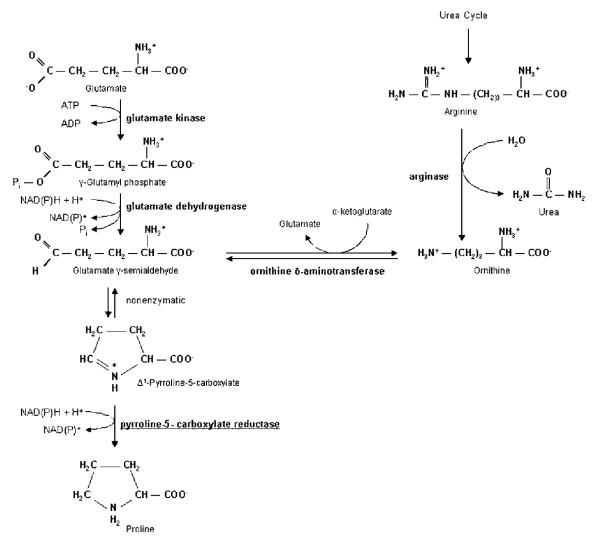
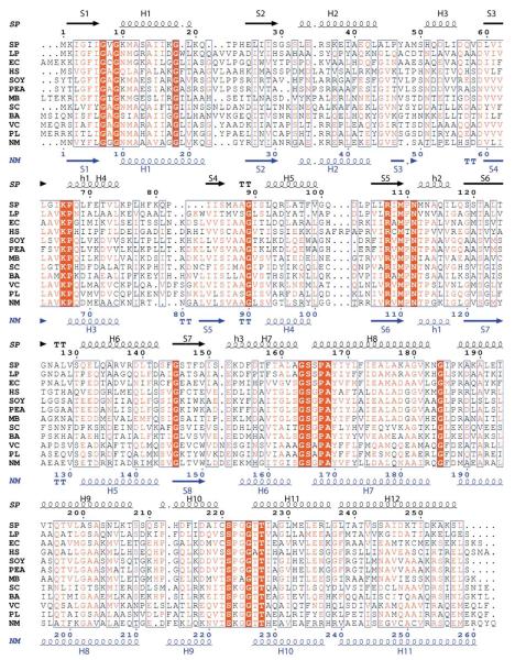
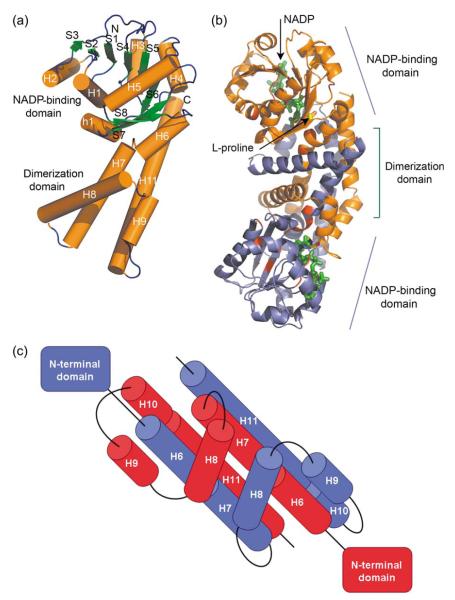
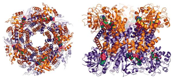
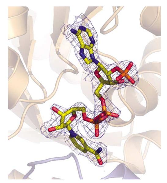
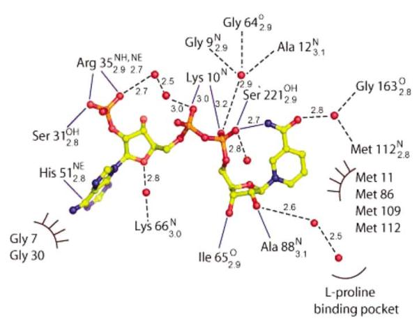
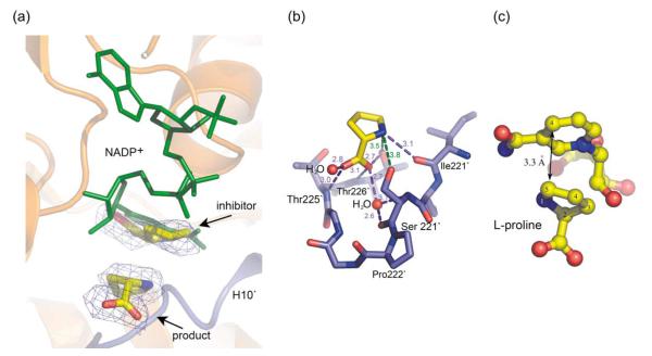
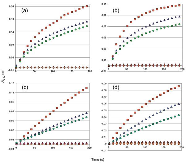
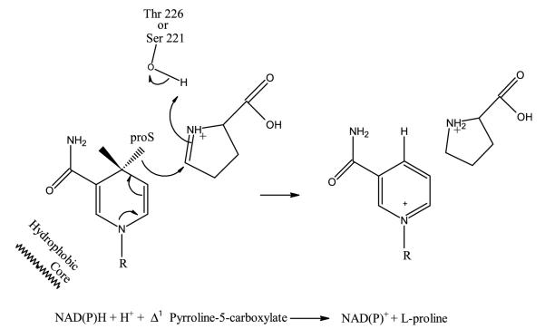
L-proline is an amino acid that plays an important role in proteins uniquely contributing to protein folding, structure, and stability, and this amino acid serves as a sequence-recognition motif. Proline biosynthesis can occur via two pathways, one from glutamate and the other from arginine. In both pathways, the last step of biosynthesis, the conversion of delta1-pyrroline-5-carboxylate (P5C) to L-proline, is catalyzed by delta1-pyrroline-5-carboxylate reductase (P5CR) using NAD(P)H as a cofactor. We have determined the first crystal structure of P5CR from two human pathogens, Neisseria meningitides and Streptococcus pyogenes, at 2.0 angstroms and 2.15 angstroms resolution, respectively. The catalytic unit of P5CR is a dimer composed of two domains, but the biological unit seems to be species-specific. The N-terminal domain of P5CR is an alpha/beta/alpha sandwich, a Rossmann fold. The C-terminal dimerization domain is rich in alpha-helices and shows domain swapping. Comparison of the native structure of P5CR to structures complexed with L-proline and NADP+ in two quite different primary sequence backgrounds provides unique information about key functional features: the active site and the catalytic mechanism. The inhibitory L-proline has been observed in the crystal structure.


Figures

Figure 1
Figure 1
Pathways of proline biosynthesis.
Figure 2
Figure 2
Multiple sequence alignment of a selected representative of the pyrroline-5-carboxylate reductase family. Sequence identities are highlighted in red and similarities are shown as red letters. The corresponding secondary structures of P5CR from S. pyogenes and N. meningitides are shown on the top (black) and the bottom (blue), respectively. Helices (H, α-helix; h, 310 helix) appear as small squiggles, beta strands (S, α-strand) as arrows. The following abbreviations were used, with Unit-Prot accession numbers indicated in parentheses: SP, Streptococcus pyogenes M1 GAS (Q9A1S9); LP, Lactobacillus plantarum (Q88Z19); EC, Escherichia coli O6 (Q8FKE0); HS, Homo sapiens (P32322); SOY, Glycine max (Soybean) (P17817); PEA, Pisum sativum (Q04708); MB, Methanococcoides burtonii; SC, Staphylococcus aureus strain COL (Q5HFR9); BA, Bacillus anthracis Ames (Q81M92); VC, Vibrio cholerae (Q9KUQ5); PL, Photorhabdus luminescens subsp. Laumondii (Q7N7H0); NM, Neisseria meningitides (Q9K1N1).
Figure 3
Figure 3
Cylinder and ribbon drawing of the P5CR dimers. (a) An overall view of the monomer of Nm-P5CR. (b) An overall view of the dimer of Sp-P5CR complexed with NADP+ (green) with the proline residues (yellow) docked into the active center. The conserved residues are highlighted in red. (c) Diagram of the C-terminal dimerization domain in Nm-P5CR.
Figure 4
Figure 4
Ribbon drawing of the dodecamer of Sp-P5CR in two views with NADP+. (a) A view down the non-crystallographic 5/2-fold relating the subunits. (b) The dodecamer viewed in a side orientation. NADP+ molecule is shown as a space-filling model in green, blue, and red.
Figure 5
Figure 5
Electron density maps (2FoFc) contoured at 1σ (blue) around NADP+ cofactor in the structure complex of Sp-P5CR/NADP+.
Figure 6
Figure 6
Schematic representation of NADP+ interactions with the surrounding protein residues (continuous blue lines) and solvent molecules (broken black line) or hydrophobic contacts (semi-circle) in molecule A of Sp-P5CR. Distances and hydrogen bond donors and acceptors are shown as subscripts and superscripts, respectively.
Figure 7
Figure 7
Diagram of sulfate binding in Nm-P5CR.
Figure 8
Figure 8
(a) Simulated annealing omit electron density maps (FoFc) contoured at 2.5σ (blue) around L-proline molecules in the structure of Sp-P5CR complexed with L-proline. The adenosine ring of the NADP+ molecule (green) overlaps with proposed inhibitory L-proline. The NADP+ molecule is shown in the orientation as observed in the Sp-P5CR/NADP+complex. (b) Diagram of the active site L-proline binding in Sp-P5CR. (c) Close-up view of the active center of superimposed NADP and proline-bound structures showing the relative positioning between hydride donor (C4-NADPH) and acceptor (C5).
Figure 9
Figure 9
Nm-P5CR ((a) and (b)) and Sp-P5CR ((c) and (d)) proline dehydrogenase activity. The enzymatic activity tested in the oxidation of L-thiazolidine-4-carboxylate (T4C) ((a) and (c)) and 3,4-dehydro-L-proline ((b) and (d)) in the presence of NADP+. The progress of the reaction was measured at room temperature at 340 mm. Reaction mixtures contained 100 mM Hepes (pH 8.0), 1 mM NADP+, 10 mM substrate T4C or 3,4-dehydro-L-proline and 0.5 μM of enzyme as a monomer in a total volume of 1 ml (red line). The orange line corresponds to reaction progress without enzyme, the brown line corresponds to reaction with 10 mM L-proline (no T4C or 3,4-dehydro-L-proline added), and the blue and green lines correspond to reactions containing 10 mM and 20 mM L-proline (with T4C or 3,4-dehydro-L-proline present).
Figure 10
Figure 10
Proposed catalytic mechanism for P5CR.

Similar articles

Cited by

References

    1. Adams E, Frank L. Metabolism of proline and the hydroxyprolines. Annu. Rev. Biochem. 1980;49:1005–1061. - PubMed
    1. Phang JM. The regulatory functions of proline and pyrroline-5-carboxylic acid. Curr. Top. Cell Regul. 1985;25:91–132. - PubMed
    1. Ambikapathy J, Marshall JS, Hocart CH, Hardham AR. The role of proline in osmoregulation in Phytophthora nicotianae. Fungal Genet. Biol. 2002;35:287–299. - PubMed
    1. Csonka LN, Hanson AD. Prokaryotic osmoregulation: genetics and physiology. Annu. Rev. Microbiol. 1991;45:569–606. - PubMed
    1. Whatmore AM, Chudek JA, Reed RH. The effects of osmotic upshock on the intracellular solute pools of Bacillus subtilis. J. Gen. Microbiol. 1990;136:2527–2535. - PubMed

Publication types

MeSH terms

Substances

LinkOut - more resources