

Human T lymphocyte responses against lung cancer induced by recombinant truncated mouse EGFR
- PMID: 16235052
- PMCID: PMC11030975
- DOI: 10.1007/s00262-005-0028-3
Human T lymphocyte responses against lung cancer induced by recombinant truncated mouse EGFR
Abstract
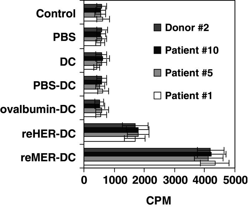
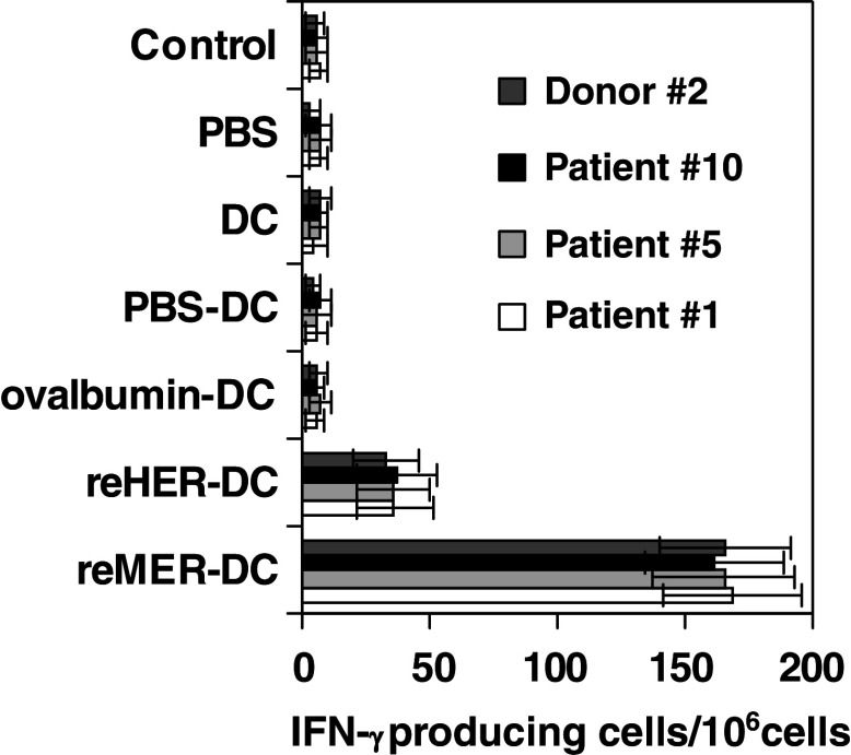
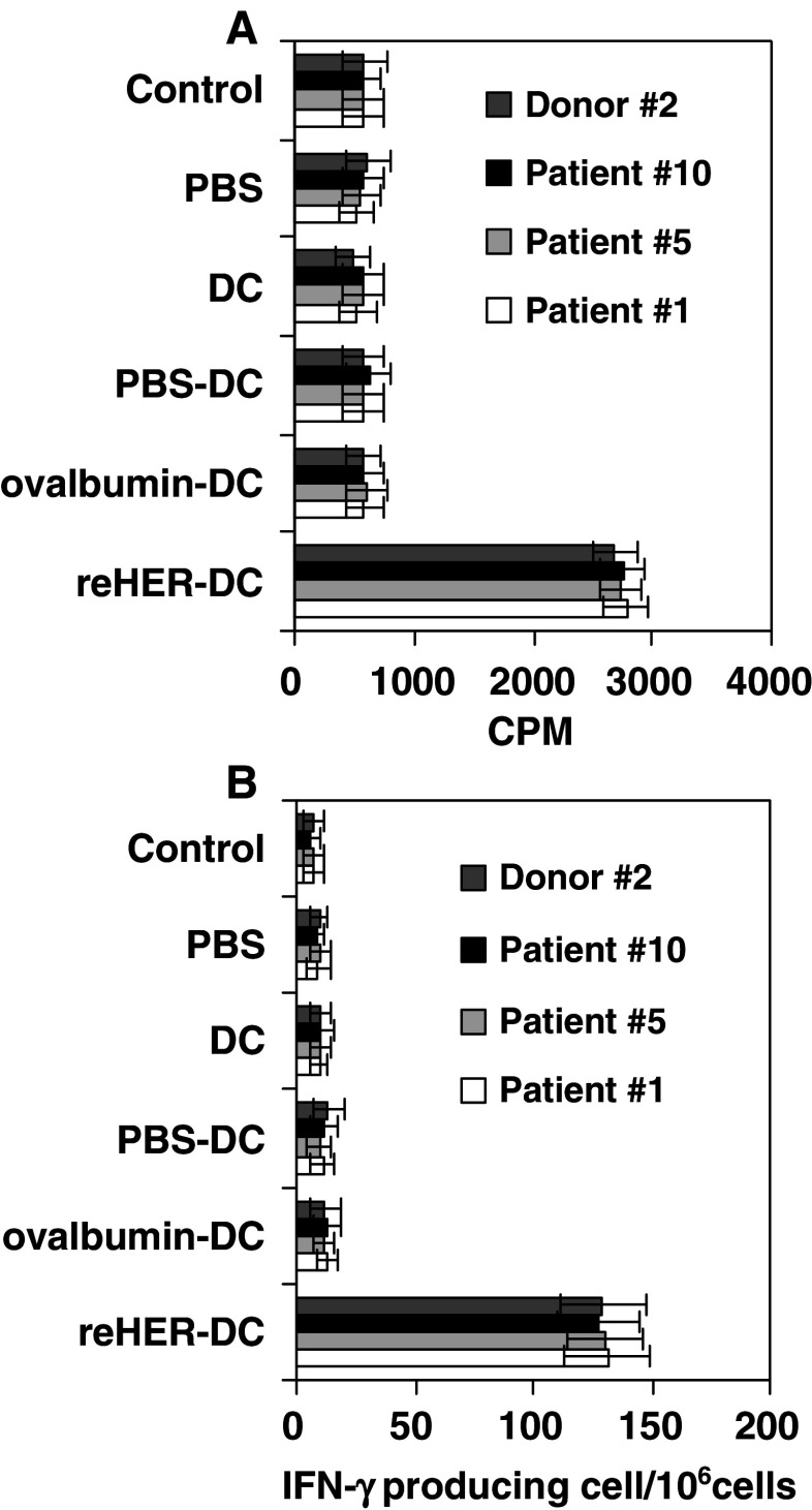
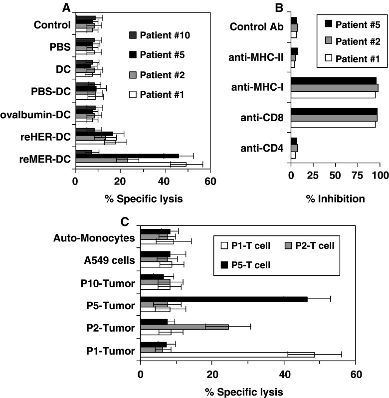
The induction of active cellular responses against EGFR should be a promising approach for the treatment of those receptor-positive tumors. However, the immunity against EGFR is presumably difficult to elicit by vaccine based on self or syngeneic EGFR due to the immune tolerance acquired during the development in immune system. We proposed a model to break immune tolerance against self-EGFR through an altered immunogen source based on xenogeneic homologous EGFR. We have previously shown human EGFR as a xenoantigen could induce specific immune responses in mouse and cross-react with mouse EGFR, and resulted in therapeutic benefits for EGFR-positive mouse tumor. Here, we show a recombinant form of extracellular domain of mouse EGFR, in the presence of DCs, could activate human peripheral T cells to proliferate, secret IFN-gamma, the induced responses could cross-react with human EGFR and kill autologous EGFR-positive lung cancer cells which could be blocked by anti-CD8 and anti-MHC class I antibody. There is no detectable cytotoxical activity against lung tissue, liver tissue and kidney tissue derived from paracancerous normal tissue. These observations suggest that antitumor immunity induced by the truncated mouse EGFR may be provoked in a cross-reaction between mouse EGFR and self-EGFR, and may provide insight into treatment of EGFR-positive tumors through induction of the autoimmune responses against EGFR.
Figures
Similar articles
-
Immunogene therapy of tumors with vaccine based on xenogeneic epidermal growth factor receptor.J Immunol. 2003 Mar 15;170(6):3162-70. doi: 10.4049/jimmunol.170.6.3162. J Immunol. 2003. PMID: 12626574
-
Active antitumor immunity elicited by vaccine based on recombinant form of epidermal growth factor receptor.J Immunother. 2005 May-Jun;28(3):236-44. doi: 10.1097/01.cji.0000161394.11831.3f. J Immunother. 2005. PMID: 15838380
-
Humoral immunity responses against EGFR-positive tumor cells induced by xenogeneic EGFR expressed in the yeast Pichia pastoris.Int J Mol Med. 2009 Feb;23(2):181-8. Int J Mol Med. 2009. PMID: 19148541
-
Xenogeneic therapeutic cancer vaccines as breakers of immune tolerance for clinical application: to use or not to use?Vaccine. 2014 Jul 7;32(32):4015-24. doi: 10.1016/j.vaccine.2014.05.006. Epub 2014 May 14. Vaccine. 2014. PMID: 24837511 Review.
-
[Tumor biotherapy based on epidermal growth factor receptor as a target].Ai Zheng. 2004 Apr;23(4):471-5. Ai Zheng. 2004. PMID: 15087042 Review. Chinese.
Cited by
-
DNA vaccines encoding altered peptide ligands for SSX2 enhance epitope-specific CD8+ T-cell immune responses.Vaccine. 2014 Mar 26;32(15):1707-15. doi: 10.1016/j.vaccine.2014.01.048. Epub 2014 Jan 31. Vaccine. 2014. PMID: 24492013 Free PMC article.
-
Prevention of human PC-346C prostate cancer growth in mice by a xenogeneic tissue vaccine.Cancer Immunol Immunother. 2007 Aug;56(8):1275-83. doi: 10.1007/s00262-006-0278-8. Epub 2007 Jan 23. Cancer Immunol Immunother. 2007. PMID: 17242926 Free PMC article.
References
Publication types
MeSH terms
Substances
LinkOut - more resources
Full Text Sources
Other Literature Sources
Medical
Research Materials
Miscellaneous
