

doi: 10.1073/pnas.0507913102.
Epub 2005 Oct 19.
Low-frequency normal mode in DNA HhaI methyltransferase and motions of residues involved in the base flipping
Affiliations
- PMID: 16236720
- PMCID: PMC1283451
- DOI: 10.1073/pnas.0507913102
Item in Clipboard
Low-frequency normal mode in DNA HhaI methyltransferase and motions of residues involved in the base flipping
Proc Natl Acad Sci U S A.
.
Abstract
The results of normal-mode analyses are in accord with the proposal that a low-frequency motion of the HhaI methyltransferase enzyme is responsible for base flipping in bound DNA. The vectors of the low-frequency normal mode of residues Ser-85 and Ile-86 point directly to the phosphate and ribose moieties of the DNA backbone near the target base in position to rotate the dihedral angles and flip the base out of the DNA duplex. The vector of residue Gln-237 on the major groove is in the proper orientation to assist base separation. Our results favor the major groove pathway and the protein active process in base flipping.
Figures
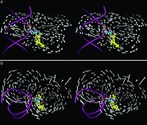
An overall picture of the low-frequency mode of the enzyme interaction. Each arrow in gray represents the direction of motion of the Cα atom. The enzyme residues are omitted for easier viewing. The DNA backbone structure, the flipped out base, and the cofactor SAM are shown in pink, yellow, and atom color, respectively. b is a top view of a. The arrows representing the large (lower right) and small (upper left) domains qualitatively form two circles. The group of arrows entering the minor groove (from the right of the helix) point directly toward the phosphodiester and sugar backbone around the flipped base. The arrows entering the major groove from the back (left) of the helix point directly to the complementary orphan guanine.
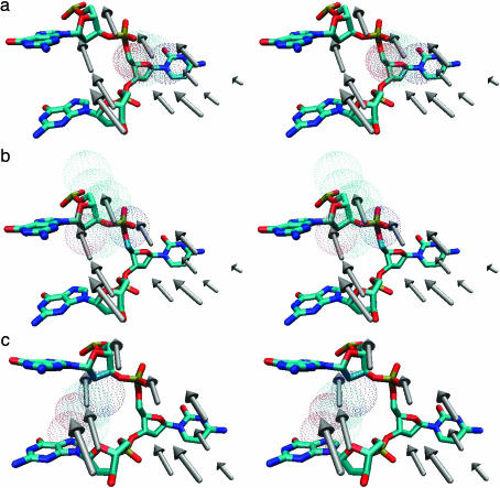
An enlarged local view of the vectors for the residues 79–89 (Phe-79, Pro-80, Cys-81, Gln-82, Ala-83, Phe-84, Ser-85, Ile-86, Ser-87, Gly-88, and Lys-89). The flipped out base and the adjacent bases of the same strand are shown explicitly. (a) Ser-85 is shown in a dotted surface fashion. The vector of this residue is located on the lower right within the top trio of vectors. (b) The vector of Ile-86, shown as a dotted surface, is located at the center and on top of the trio of vectors. (c) The Ser-87 vector, shown in a dotted surface fashion, is located on the lower left of the top trio of vectors. Ser-87 is situated on the minor groove side of the DNA between the adjacent bases. The Ser-87 vector also points to the sugar of the adjacent guanine on the 5′ side.
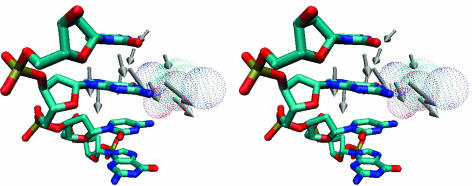
An enlarged local view of the vectors for the residues 230–240 (Gly-230, Ile-231, Val-232, Gly-233, Lys-234, Gly-235, Gly-236, Gln-237, Gly-238, Glu-239, and Arg-240). These vectors are located in the back of the major groove of the small domain and point toward the complementary orphan guanine base (Fig. 1 a and b). The orphan guanine and the adjacent bases of the same strand are shown explicitly. Residue Gln-237, shown in a dotted surface fashion, is situated on the major groove side in front of the group of vectors.
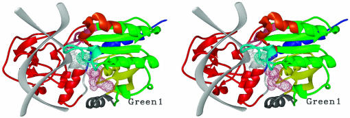
Anticorrelated motions mapped onto the 3D stereo DNA methyltransferase M.HhaI in a complex [d(CCATGCGCTGAC)2 and AdoMet]. The DNA backbone is shown as a light gray ribbon. The flipped out base and AdoMet are shown by light gray and pink dotted surfaces, respectively. The Cys-81 side chain is shown as a dotted cyan surface. Different anticorrelated regions are color coded for easy viewing. Residues 186–279 and 291–304 (red) of the small domain are anticorrelated with residues 1–40 (yellow), 55–79, 98–120, 135–150, and 165–185 (green) and 305–327 (blue) in large catalytic domain. Residues 280–290 (light gray) of the small domain are anticorrelated with residues 80–97 (cyan) in the large catalytic domain. Residues 80–97 are also anticorrelated, within the large catalytic domain, with residues 1–40 (yellow) and 305–327 (blue). The following anticorrelated motions are also within the large catalytic domain: residues 121–134 (orange) are anticorrelated with residues 1–40 (yellow) and residues 41–54 (gray). The pink region, residues 151–164, is anticorrelated with residues 41–54 (gray) and residues 55–79 (labeled green 1).
Similar articles
-
Caught in the act: visualization of an intermediate in the DNA base-flipping pathway induced by HhaI methyltransferase.Nucleic Acids Res. 2004 Jul 23;32(13):3877-86. doi: 10.1093/nar/gkh701. Print 2004. Nucleic Acids Res. 2004. PMID: 15273274 Free PMC article.
-
Protein-facilitated base flipping in DNA by cytosine-5-methyltransferase.Proc Natl Acad Sci U S A. 2003 Jan 7;100(1):68-73. doi: 10.1073/pnas.0135427100. Epub 2002 Dec 27. Proc Natl Acad Sci U S A. 2003. PMID: 12506195 Free PMC article.
-
Structures of HhaI methyltransferase complexed with substrates containing mismatches at the target base.Nat Struct Biol. 1998 Oct;5(10):872-7. doi: 10.1038/2312. Nat Struct Biol. 1998. PMID: 9783745
-
Finding a basis for flipping bases.Structure. 1996 Jun 15;4(6):639-45. doi: 10.1016/s0969-2126(96)00068-8. Structure. 1996. PMID: 8805547 Review.
-
Atomistic view of base flipping in DNA.Philos Trans A Math Phys Eng Sci. 2004 Jul 15;362(1820):1439-60. doi: 10.1098/rsta.2004.1383. Philos Trans A Math Phys Eng Sci. 2004. PMID: 15306460 Review.
Cited by
-
Base Dynamics in the HhaI Protein Binding Site.J Phys Chem B. 2023 Aug 24;127(33):7266-7275. doi: 10.1021/acs.jpcb.3c03687. Epub 2023 Aug 10. J Phys Chem B. 2023. PMID: 37561575 Free PMC article.
-
Hidden Conformation Events in DNA Base Extrusions: A Generalized Ensemble Path Optimization and Equilibrium Simulation Study.J Chem Theory Comput. 2013 Aug 13;9(8):10.1021/ct400198q. doi: 10.1021/ct400198q. J Chem Theory Comput. 2013. PMID: 24250279 Free PMC article.
-
A molecular dynamics study of slow base flipping in DNA using conformational flooding.Biophys J. 2007 Aug 1;93(3):770-86. doi: 10.1529/biophysj.106.091751. Epub 2007 May 11. Biophys J. 2007. PMID: 17496048 Free PMC article.
-
Coupling between catalytic loop motions and enzyme global dynamics.PLoS Comput Biol. 2012;8(9):e1002705. doi: 10.1371/journal.pcbi.1002705. Epub 2012 Sep 27. PLoS Comput Biol. 2012. PMID: 23028297 Free PMC article.
References
-
- Doerfler, W. (1983) Annu. Rev. Biochem. 52, 93–124. - PubMed
-
- Wu, J. C. & Santi, D. V. (1987) J. Biol. Chem. 262, 4778–4786. - PubMed
-
- Roberts, R. J., Myers, P. A., Morrison, A. & Murray, K. (1976) J. Mol. Biol. 103, 199–208. - PubMed
-
- Mann, M. B. & Smith, H. O. (1979) in Proceedings of the Conference on Transmethylation, eds. Usdin, E., Borchardt, R. T. & Creveling, C. R. (Elsevier, New York), pp. 483–492.
Publication types
MeSH terms
Substances
Grants and funding
LinkOut - more resources
Full Text Sources
Molecular Biology Databases
