. 2005 Nov 1;102(44):15971-6.
doi: 10.1073/pnas.0503868102. Epub 2005 Oct 18.

Combining two genomes in one cell: stable cloning of the Synechocystis PCC6803 genome in the Bacillus subtilis 168 genome

Affiliations

Combining two genomes in one cell: stable cloning of the Synechocystis PCC6803 genome in the Bacillus subtilis 168 genome

Mitsuhiro Itaya et al. Proc Natl Acad Sci U S A. .

Abstract

Cloning the whole 3.5-megabase (Mb) genome of the photosynthetic bacterium Synechocystis PCC6803 into the 4.2-Mb genome of the mesophilic bacterium Bacillus subtilis 168 resulted in a 7.7-Mb composite genome. We succeeded in such unprecedented large-size cloning by progressively assembling and editing contiguous DNA regions that cover the entire Synechocystis genome. The strain containing the two sets of genome grew only in the B. subtilis culture medium where all of the cloning procedures were carried out. The high structural stability of the cloned Synechocystis genome was closely associated with the symmetry of the bacterial genome structure of the DNA replication origin (oriC) and its termination (terC) and the exclusivity of Synechocystis ribosomal RNA operon genes (rrnA and rrnB). Given the significant diversity in genome structure observed upon horizontal DNA transfer in nature, our stable laboratory-generated composite genome raised fundamental questions concerning two complete genomes in one cell. Our megasize DNA cloning method, designated megacloning, may be generally applicable to other genomes or genome loci of free-living organisms.


Figures

Fig. 1.
Fig. 1.
IWe steps to megacloning. A set of LPS was prepared by PCR and arranged in tandem in pBR322-derived vector in E. coli to yield a set of LPA plasmids. LPA plasmids, constructed from pCISP310B and pCISP311B (12) or pCISP334 and pCISP335 (21), have either a cat (filled circle) or an erm (open circle) marker used alternatively. The LPA[1/2] plasmid was delivered to the GpBR sequence by means of double-homologous recombination as indicated by X. IWe[1/2] indicates that Synechocystis DNA taken up by competent B. subtilis cells recombines at LPA and the internal continuous green-colored sequence integrates. An LPA[2/3] plasmid is delivered to conduct the next IWe[2/3]. Repeats of LPA[n/n + 1] and IWe[n/n + 1] elongate the continuous Synechocystis genome DNA in GpBR. Delivery of LPA was selected by cat or erm alternatively. IWe selection is by an internal positive selection system (13) briefly described in Methods. The heavy gold arrow in LPA indicates that the λ cI857 repressor gene linked with the spectinomycin resistance gene (spc) was repeatedly used. Strains for all IWe are described in Fig. 6. I-PpoI sites at both ends of GpBR, indicated by vertical lines inside BGM at the bottom, always remain unchanged and were used to produce cloned DNA in GpBR as shown in Fig. 4.
Fig. 2.
Fig. 2.
Structure of the megacloned Synechocystis genome in the B. subtilis genome. Schematic drawings of the genomes of Synechocystis PCC6803 (green circle) (17), B. subtilis 168 (brown circle), and strain BEST7613 (mosaic with green and brown). Seven plasmids reported for Synechocystis are omitted. B. subtiis 168 has no indigenous plasmid. Regions A, B, and C are from the B. subtilis genome and regions [I]-[IV] are from the Synechocystis genome. The BEST7613 genome (7,700 kb) shows approximate size because the B. subtilis strain used in this study, BEST7003 (13), had certain deletions and was shorter than the reported strain (26). The locations of oriC and terC are known for B. subtilis only. Antibiotic resistance markers inserted in the BEST7613 genome are shown with relevant genes and locations: resistance to blasticidin S (bsr), tetracycline (tet), spectinomycin (spc), neomycin (neo), and phleomycin (phl). The GpBR (indicated by the striped boxes) is the megacloning locus prepared at the proB, leuB and ytqB loci of the B. subtilis genome. I-PpoI sites indicated by bars inside the BEST7003 and BEST7163 circle are created at the ends of GpBRs and of three megacloned Synechocystis genomes. The GpBR sequences that remained in BEST7613 after removal manipulations are shown. GpBR between regions A and [I] was removed after BEST7155 (Fig. 3). The GpBR sequence between regions [II] and B was replaced by tet before BEST7566 (Fig. 3) to reduce the overlap of regions [II] and [III]. GpBR between regions B and [IV] was replaced by spc before BEST7374 (Fig. 3). The I-PpoI site is left behind upon all of these removals. Overlaps of 5.984 kb for regions [II] and [III], 4.479 kb for regions [III] and [IV], and 0.334 kb for regions [IV] and [I] remained in BEST7613. Six fragments yielded from BEST7613 by I-PpoI digestion are shown in Fig. 4.
Fig. 3.
Fig. 3.
All of the steps to complete megacloning of the Synecohsystis genome. (Upper Left and Upper Center) Four Synechocystis genome regions (Upper Left) with sll1652 (filled oval; open oval if deleted) and rrn genes (filled box, open box if deleted) were megacloned by IWe (Upper Center). (Right) The four megacloned recombinants were used to complete BEST7613 by IWeT; oval and box symbols correspond to those for Upper Left and Upper Center. For Upper Center and Right, the two short bars connected to the outside of the genome circle represent oriC (top) and terC (bottom), schematically indicating genome symmetry. (Lower Center) BGM vectors are schematically drawn with the GpBR at three loci. (Lower Left) Colonies formed on LB plates during 17 h at 37°C.
Fig. 4.
Fig. 4.
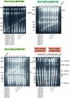
IWe and IWeT steps in megacloning. I-PpoI fragments from indicated BGM strains run on CHEF gels with size markers indicated to the right. (Upper Left) I-PpoI fragments of cloned region [I] indicated by green arrowheads. (Upper Right) Strains of IWeT-mediated integration and elongation of region [IV] in BEST7324 (BEST7155 plus sll1652 disrupted by bsr) to BEST7374. (Lower Left) Strains of IWeT-mediated integration and elongation of region [II] in BEST7527 to BEST7566. (Lower Right) IWeT-mediated integration and elongation of region [III]. Indicated strains of IWeT of region [III] are separated into preintegration (lanes 1-4) and postintegration (lanes 5-9) of region [II]. Synechocystis genome DNA and the BGM vector part are indicated by differently colored arrowheads. Six I-PpoI fragments produced from BEST7613 are mapped to those in Fig. 2. Running conditions are shown. Yeast chromosomal DNA was run as a size marker.
Fig. 5.
Fig. 5.
IWeT steps for assembly of the megacloned Synechocystis genome segment in BGM. IWeT of region [IV] to the leuB locus of the strain BEST7324 designated in the legend to Fig. 4 Upper Right; region [I] at proB is shown as an example. Selected IWe strains indicated by orange and green arrows from IWe region [IV] shown in Fig. 6 were used. The integration started by IWeT[1/2] is followed by alternate use of end markers indicated by cat (filled circle) or erm (open circle). The supply of longer regions for homologous recombination (indicated by a blue X) resulted in more effective elongation than was possible with IWe.

References

    1. Doolittle, R. F. (1998) Nature 392, 339-342. - PubMed
    1. Jain, R., Rivera, M. C. & Lake, J. A. (1999) Proc. Natl. Acad. Sci. USA 96, 3801-3806. - PMC - PubMed
    1. Gophna, U., Charlebois, R. L. & Doolittle, W. F. (2004) Trends Microbiol. 12, 213-219. - PubMed
    1. Heidelberg, J. F., Eisen, J. A., Nelson, W. C., Clayton, R. A., Gwinn, M. L., Dodson, R. J., Haft, D. H., Hickey, E. K., Peterson, J. D., Umayam, L, et al. (2000) Nature 406, 477-483. - PMC - PubMed
    1. White, O., Eisen, J. A., Heidelberg, J. F., Hickey, E. K., Peterson, J. D., Dodson, R. J., Haft, D. H., Gwinn, M. L., Nelson, W. C., Richardson, D. L., et al. (1999) Science 286, 1571-1577. - PMC - PubMed