. 2006 Jan;17(1):511-24.
doi: 10.1091/mbc.e05-07-0682. Epub 2005 Oct 19.

GMx33 associates with the trans-Golgi matrix in a dynamic manner and sorts within tubules exiting the Golgi

Affiliations

GMx33 associates with the trans-Golgi matrix in a dynamic manner and sorts within tubules exiting the Golgi

Christopher M Snyder et al. Mol Biol Cell. 2006 Jan.

Abstract

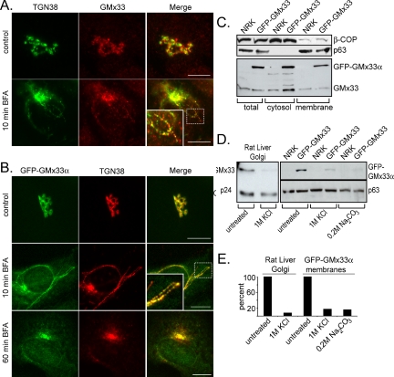
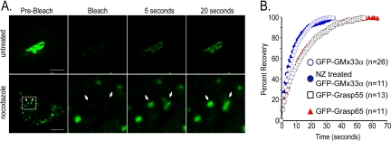
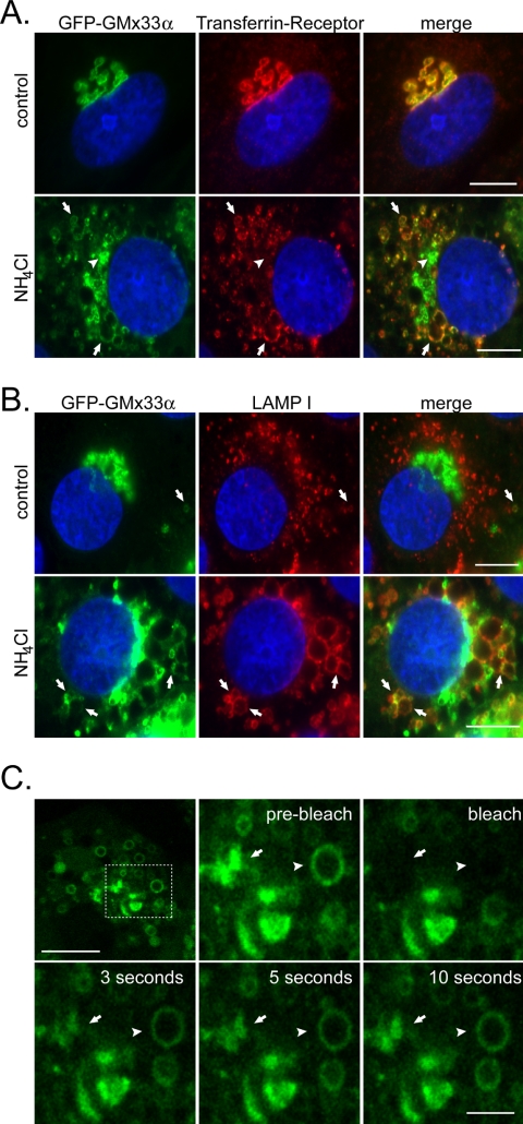
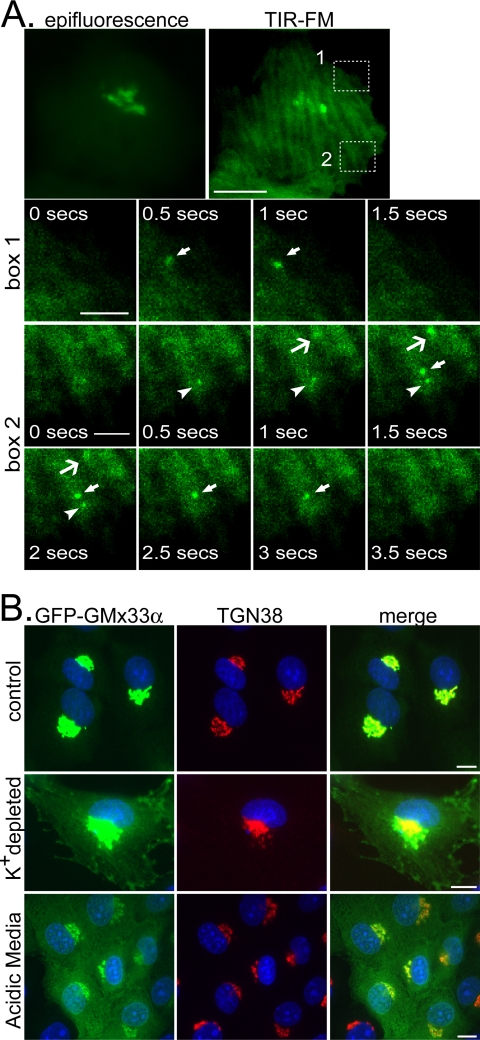
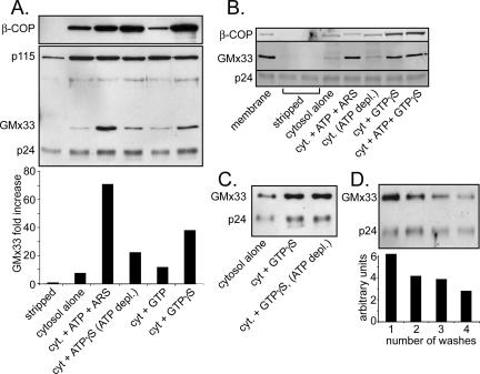
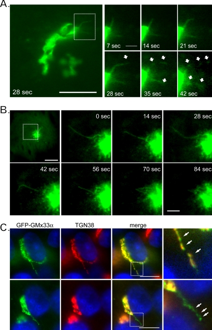
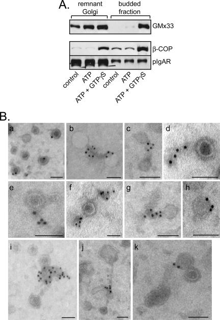
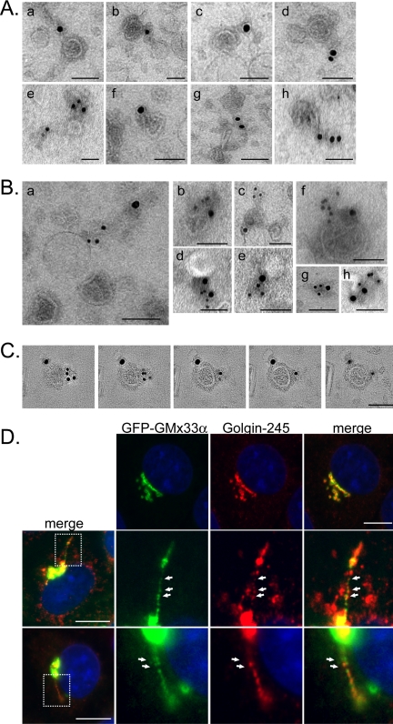
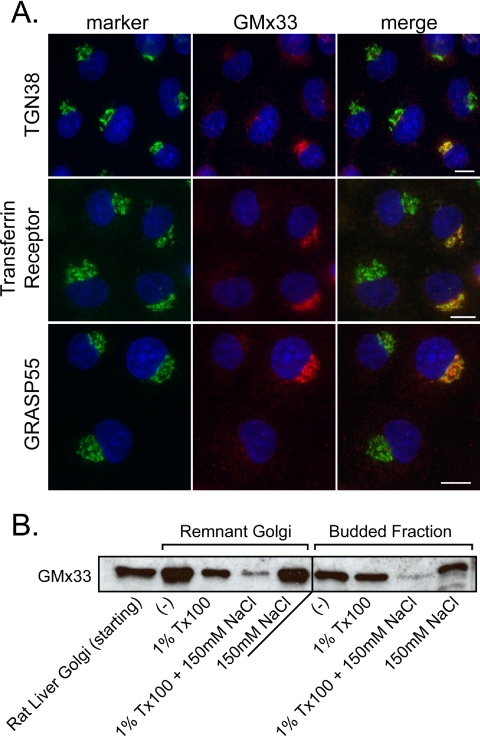
The trans-Golgi matrix consists of a group of proteins dynamically associated with the trans-Golgi and thought to be involved in anterograde and retrograde Golgi traffic, as well as interactions with the cytoskeleton and maintenance of the Golgi structure. GMx33 is localized to the cytoplasmic face of the trans-Golgi and is also present in a large cytoplasmic pool. Here we demonstrate that GMx33 is dynamically associated with the trans-Golgi matrix, associating and dissociating with the Golgi in seconds. GMx33 can be locked onto the trans-Golgi matrix by GTPgammaS, indicating that its association is regulated in a GTP-dependent manner like several other Golgi matrix proteins. Using live-cell imaging we show that GMx33 exits the Golgi associated with tubules and within these tubules GMx33 segregates from transmembrane proteins followed by fragmentation of the tubules into smaller tubules and vesicles. Within vesicles produced by an in vitro budding reaction, GMx33 remains segregated in a matrixlike tail region that sometimes contains Golgin-245. This trans-matrix often links a few vesicles together. Together these data suggest that GMx33 is a member of the trans-Golgi matrix and offer clues regarding the role of the trans-Golgi matrix in sorting and exit from the Golgi.


Figures

Figure 1.
Figure 1.
Endogenous GMx33 and GFP-GMx33α are peripherally localized to the TGN. (A) NRK cells were left untreated (top panels) and treated with 5 μg/ml brefeldin A for 10 min (bottom panels), fixed, and stained with antibodies specific for either TGN38 or GMx33. Our GMx33-specific antibody recognizes both α and β forms of GMx33. Primary antibodies were detected with either anti-mouse or anti-rabbit secondary antibodies conjugated to either Alexa Fluor 488 or Alexa Fluor 594. The inset is an enlarged image from the area indicated by the dotted box. (B) NRK cells stably expressing GFP-GMx33α were left untreated (top panel) or treated with brefeldin A for 10 or 60 min (middle and bottom panels). Fixed cells were stained for TGN38. The inset is an enlarged image from the area indicated by the dotted box. (C) NRK cells stably expressing GFP-GMx33α were lysed in detergent (total) or used to prepare membrane and cytosol fractions as described in Materials and Methods. Protein (50 μg per lane) was analyzed by immunoblot with antibodies specific for the indicated proteins. (D) Fifteen micrograms of rat liver Golgi fraction were left untreated or stripped with 1 M KCl for 1 h on ice and washed (left panel). One hundred micrograms of total membrane fractions from wild-type NRK cells or those expressing GFP-GMx33α (right panel) were left untreated or stripped with 1 M KCl as above or a high pH wash, 0.2 M sodium carbonate for 15 min on ice. Half of each sample was subjected to SDS-PAGE and analyzed by immunoblot with antibodies specific for the indicated proteins. p24 and p63 proteins were followed as loading controls. (E) Densitometry was performed on the immunoblots in D using the NIH Image J software. The amount of p24 or p63 signal in each lane was used to normalize slight differences in loading. Bars, 10 μm.
Figure 2.
Figure 2.
GFP-GMx33α rapidly cycles between membrane and cytosolic pools. (A) Cells stably or transiently expressing GFP-GMx33α were left untreated (top panels) or were treated with 33.3 μM nocodazole for 2 h at 37°C (bottom panels). The whole Golgi region or the indicated Golgi-stacklets were bleached with the 488-nm laser set to 100%. Approximately one image was captured each second after bleaching and the fluorescence recovery at 5 and 20 s is shown. The bottom leftmost panel shows the entire cell. The region indicated by the white dotted box is enlarged in the right three panels. (B) The indicated number of cells transfected with GFP-GMx33α, GFP-GRASP55, or GFP-GRASP65 were bleached and the average fluorescence recovery after bleaching was graphed. The actual fluorescence intensity for each cell over time was converted to a percentage of the total recovery. Fluorescence values obtained immediately after bleaching were used as 0%. One hundred percent recovery corresponds to the point for each cell at which overall fluorescence no longer increased over time. Because of the rapid exchange of membrane and cytosolic forms of GFP-GMx33α, the cytosolic pool was bleached as it exchanged on the membrane during the bleaching period. Therefore, the total fluorescence never recovered to the initial prebleach levels, and recovery level was inversely proportional to the time of bleaching. Bars, 10 μm.
Figure 3.
Figure 3.
GMx33 can be “trapped” at endosomal membranes. (A) NRK cells stably transfected with GFP-GMx33α were left untreated or treated with ammonium chloride for 2 h before fixation and staining. Cells were stained with an antibody specific for the transferrin receptor and DAPI. Arrows indicate enlarged endosomal membranes positive for both GFP-GMx33α and the transferrin receptor. Arrowheads indicate GFP-GMx33α that is still associated with the Golgi. Bar, 10 μm. (B) Cells were treated as in A and stained with an antibody specific for LAMP I and DAPI. Arrows indicate LAMP I-positive membranes that are also positive for GFP-GMx33α in both control cells (top panels) and ammonium chloride treated (bottom panels) cells. Bar, 10 μm. (C) NRK cells stably transfected with GFP-GMx33α were treated with ammonium chloride as in A and B. An enlarged endosome (arrowhead) and an edge of the Golgi (arrow) were both bleached as in Figure 2. The dotted box in the leftmost top panel indicates the region enlarged in the other panels. Bar in top leftmost panel, 10 μm. Bar in bottom right panel, 3 μm.
Figure 4.
Figure 4.
GFP-GMx33α can be found at the plasma membrane by TIR-FM and can be trapped at the plasma membrane by inhibitors of endocytosis. (A) NRK cells stably transfected with GFP-GMx33α were maintained at 37°C and visualized by TIR-FM over time. The epifluorescent image of the entire cell is also shown (top left panel). The bottom three rows show enlarged images of the boxed areas in the top right-hand panel. Images were captured every 0.5 s. Arrows and arrowheads point to GFP-GMx33α-positive events at the plasma membrane. This localization is transient. These events are seen more clearly in Supplementary Video 1. Bar in top panels, 10 μm; bars in bottom panels, 3 μm. (B) NRK cells stably transfected with GFP-GMx33α were left untreated or were cultured in the presence of a K+-depleting buffer or acidic (pH 5) media. Cells were fixed and stained with antibodies specific for TGN38 before imaging. Bars, 10 μm.
Figure 5.
Figure 5.
Cytosolic GMx33 associates with stripped membranes in a GTPor ATP-dependent manner. (A) Rat liver Golgi fraction was stripped with 1 M KCl, split into 15 μg aliquots, and incubated with cytosol (800 μg per aliquot) for 30 min with the indicated treatment as described in Materials and Methods. Half of each sample was analyzed by immunoblot with antibodies against the following proteins: β-COP, p115, GMx33, and p24. For densitometry, the GMx33 signal was standardized to the signal for the Golgi-transmembrane protein, p24, and the data are in the bottom part of the panel. (B) Rat liver Golgi fraction was stripped with 2 M NaCl. Cytosolic proteins were incubated with the stripped membrane in the presence of the indicated nucleotides and samples were analyzed as in A. (C) Rat liver Golgi fraction was stripped as in A and treated as indicated with the indicated nucleotides. (D) Rat liver Golgi fraction was not stripped and was subjected to the indicated number of washes at 4°C. Between each wash the fraction was incubated in cytosol buffer for 10 min at 37°C. The final membrane pellets were immunoblotted with antibodies specific for GMx33 and p24. Quantitation was performed as in A and is provided below the blot.
Figure 6.
Figure 6.
GFP-GMx33α exits the Golgi in tubules. (A) NRK cells stably expressing GFP-GMx33α were held at 37°C and visualized by time-lapse imaging every 7 s. Shown is a pair of tubules extending from the Golgi that fragment into short tubules or vesicles between 28 and 42 s. The left panel shows the whole Golgi; the right panels are enlarged images of the region indicated by a dashed box. Bar in leftmost panel, 10 μm; in enlarged images, 2 μm. (B) NRK cells stably expressing GFP-GMx33α were treated with brefeldin A for 1 h and visualized by time-lapse imaging as in A. Shown is a whole cell just before tubule formation and enlarged images of the region outlined by the dotted box in the top leftmost panel. Bar in the leftmost top panel, 10 μm; in the enlarged images, 3μm. (C) NRK cells stably transfected with GFP-GMx33α were fixed and stained with antibodies specific for TGN38. Two cells with tubules extending from the Golgi are shown. Bars, 10 μm.
Figure 7.
Figure 7.
GMx33 associates in a GTP-dependent manner with matrixlike structures attached to vesicles in a budded vesicle fraction. (A) Rat liver Golgi fraction (75 μg) was incubated with 4.5 mg cytosol and the indicated nucleotides for 30 min at 37°C and remnant Golgi and budded fractions were separated as described in Materials and Methods. Protein was analyzed by immunoblot with antibodies specific for GMx33, β-COP, and pIgA receptor (a cargo molecule moving to the plasma membrane). (B) Budded fractions were prepared as in A in the presence of ATP and GTPγS, placed on an EM grid, and immunolabeled with antibodies specific for GMx33. Shown are 11 different examples of budded vesicles associated with electron dense (osmophilic) “tails” that are positive for GMx33. Some of these tails appear to connect adjacent vesicles (b, c, g, j, and k). The gold particles lie almost exclusively within the tail material and are distinct from the vesicles' membranes. Bars, 100 nm.
Figure 8.
Figure 8.
GMx33 associates with the trans- Golgi matrix and colocalizes with p230/Golgin-245 in tubules exiting the Golgi. (A) Budded fractions were immunolabeled on EM grids as in Figure 7 with antibodies specific for p230/Golgin-245. Shown are eight different examples of Golgin-245-positive electron dense (osmophilic) “tails” associated with the budded vesicles. Bars, 100 nm. (B) Budded fractions were prepared as in A and probed with antibodies specific for p230/Golgin-245 (15 nm gold) and GMx33 (10 nm gold). Shown are eight examples of structures, both vesicles and matrix alone (g) that colabeled for both proteins. Bars, 100 nm. (C) A vesicle that colabeled with antibodies for GMx33 and Golgin-245 was analyzed by 3-D tomography. Shown are five tomographic slices (0.45 nm) through the volume of the vesicle. Gold particles corresponding to the two antigens are localized to the electron-dense matrix material surrounding the vesicle and both are distinct from the vesicle membrane itself. Bar, 100 nm. (D) NRK cells stably transfected with GFP-GMx33α were fixed and labeled with antibodies specific for p230/Golgin-245 and fluorophore-conjugated secondary antibodies. The top panels show that there is good, but not perfect colocalization between GMx33α and Golgin-245. The bottom two panels show a tubule extending from different cells. The boxed regions in the leftmost panels depict the areas enlarged in the subsequent panels. Bars, 10 μm.
Figure 9.
Figure 9.
Depletion of GMx33 does not visibly disrupt the Golgi structure even though GMx33 has the biochemical properties of the Golgi matrix. (A) NRK cells were transfected with siRNA specific for GMx33α, incubated for 96 h, and stained with antibodies specific for GMx33 and the indicated marker protein. Each image includes cells that continued to express GMx33 for comparison. Bar, 10 μm. (B) Budded fractions were isolated from 75 μg rat liver Golgi fraction in the presence of ATP and GTPγS. After isolation, the pelleted budded fractions or remnant Golgi fractions were analyzed directly (-) subjected to extraction with 1% Tx100 alone, 1% Tx100 + 150 mM NaCl, or 150 mM NaCl alone for 30 min on ice. Each sample was then pelleted again and protein was analyzed by immunoblot with antibodies specific for GMx33.

References

    1. Allan, V. J., and Kreis, T. E. (1986). A microtubule-binding protein associated with membranes of the Golgi apparatus. J. Cell Biol. 103, 2229-2239. - PMC - PubMed
    1. Amos, W. B., and Grimstone, A. V. (1968). Intercisternal material in the Golgi body of Trichomonas. J. Cell Biol. 38, 466-471. - PMC - PubMed
    1. Barr, F. A., Nakamura, N., and Warren, G. (1998). Mapping the interaction between GRASP65 and GM130, components of a protein complex involved in the stacking of Golgi cisternae. EMBO J. 17, 3258-3268. - PMC - PubMed
    1. Barr, F. A., Puype, M., Vandekerckhove, J., and Warren, G. (1997). GRASP65, a protein involved in the stacking of Golgi cisternae. Cell 91, 253-262. - PubMed
    1. Barr, F. A., and Short, B. (2003). Golgins in the structure and dynamics of the Golgi apparatus. Curr. Opin. Cell Biol. 15, 405-413. - PubMed

Publication types

MeSH terms

LinkOut - more resources