. 2006 Jan;17(1):32-42.
doi: 10.1091/mbc.e05-04-0339. Epub 2005 Oct 19.

Chironomus tentans-repressor splicing factor represses SR protein function locally on pre-mRNA exons and is displaced at correct splice sites

Affiliations

Chironomus tentans-repressor splicing factor represses SR protein function locally on pre-mRNA exons and is displaced at correct splice sites

Petra Björk et al. Mol Biol Cell. 2006 Jan.

Abstract

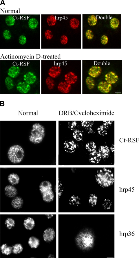
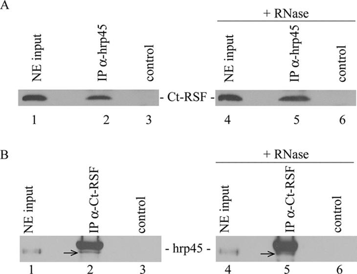
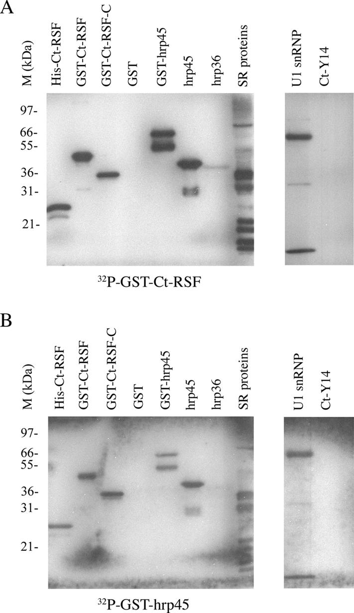
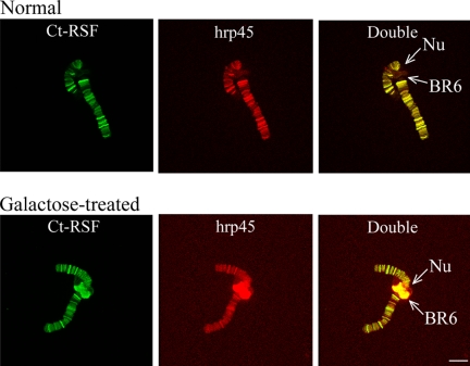
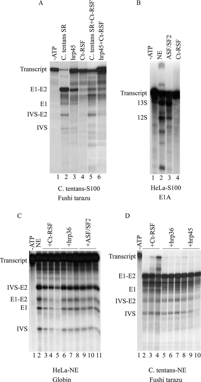
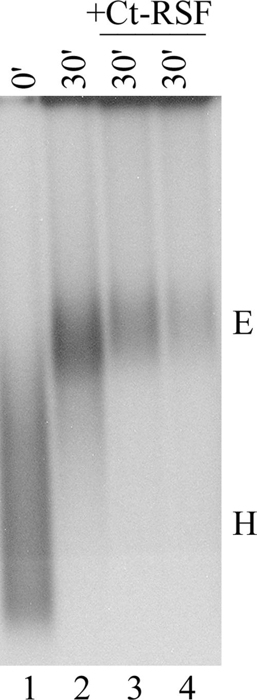
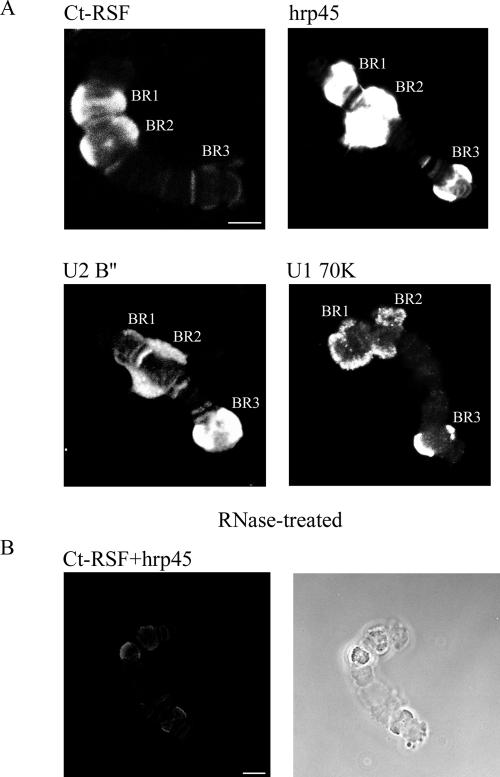
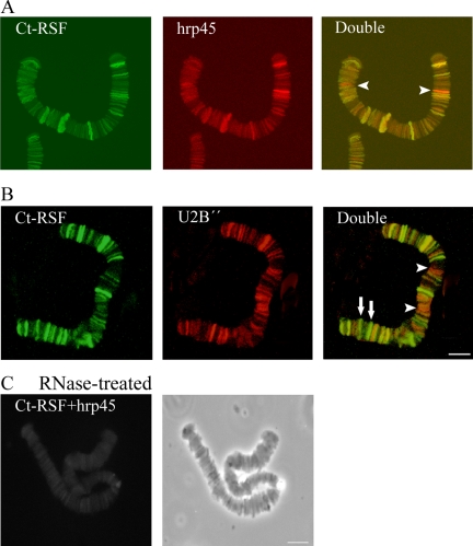
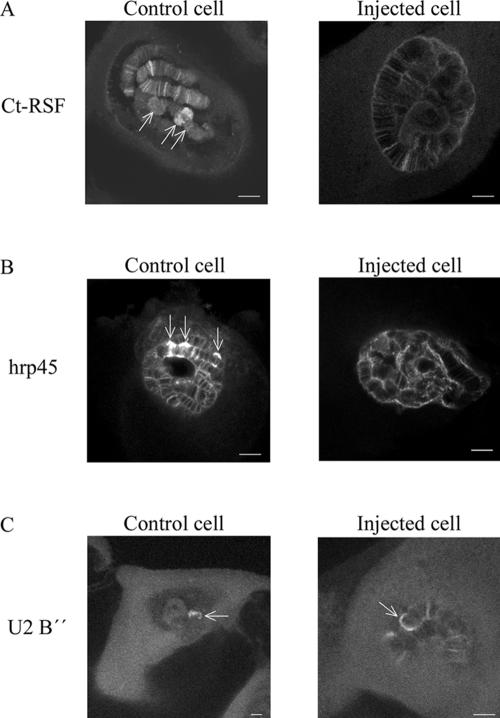
Chironomus tentans-repressor splicing factor (Ct-RSF) represses the activation of splicing by SR proteins in vitro. Ct-RSF colocalizes with the Ser-Arg-rich (SR) protein hrp45 in interchromatin granule clusters and coimmunoprecipitates with hrp45 in nuclear extracts. Ct-RSF and hrp45 can also interact directly in vitro. Ct-RSF and hrp45 are recruited together to transcribing genes and associate with growing pre-mRNAs. Ct-RSF and hrp45 colocalize at a large number of gene loci. Injection of anti-Ct-RSF antibodies into nuclei of living cells blocks association of both Ct-RSF and hrp45 with the growing pre-mRNA, whereas binding of U2 small nuclear ribonucleoprotein particle (snRNP) to the pre-mRNA is unaffected. On the intron-rich Balbiani ring (BR) 3 pre-mRNA, hrp45 as well as U1 and U2 snRNPs bind extensively, whereas relatively little Ct-RSF is present. In contrast, the BR1 and BR2 pre-mRNAs, dominated by exon sequences, bind relatively much Ct-RSF compared with hrp45 and snRNPs. Our data suggest that Ct-RSF represses SR protein function at exons and that the assembly of spliceosomes at authentic splice sites displaces Ct-RSF locally.


Figures

Figure 1.
Figure 1.
Ct-RSF is restricted to the nucleus and colocalizes with the SR protein hrp45 in nuclear speckles. (A) C. tentans diploid cells were stained with anti-Ct-RSF and anti-hrp45 antibodies. In cells grown in normal medium (top), the staining patterns for the two proteins overlapped extensively. In cells treated with actinomycin D (bottom), both Ct-RSF and hrp45 were redistributed into the same larger and more intensively stained speckles. (B) Ct-RSF and hrp45 were restricted to the nucleus and shifted into large speckles also upon treatment with DRB and cycloheximide. In contrast, the hnRNP protein hrp36 did not accumulate in the same speckles. To some extent, it accumulated in the cytoplasm. Bars, 2 μm.
Figure 2.
Figure 2.
Ct-RSF and hrp45 are coimmunoprecipitated. Nuclear extract from C. tentans cells was immunoprecipitated with anti-hrp45 antibodies (A) or anti-Ct-RSF antibodies (B). Ct-RSF coimmunoprecipitated with hrp45 and vice versa (A and B, lanes 2). The hrp45 signal in B (arrows) is located just below the position of the antibody used in the experiment. Controls represent immunoprecipitations in the absence of specific antibodies. RNase treatment of the nuclear extracts before immunoprecipitation did not affect the signal (A and B, lanes 5).
Figure 3.
Figure 3.
Ct-RSF and hrp45 bind directly to each other in vitro. Far-Western experiment in which the indicated proteins were separated on polyacrylamide gels, blotted to a nylon filter, and refolded. The proteins were probed with labeled Ct-RSF (A) or labeled hrp45 (B). SR proteins were purified from C. tentans cells. U1 snRNP represents purified mammalian U1 snRNP. The positions of size marker proteins are shown to the left of each filter.
Figure 4.
Figure 4.
t-RSF and hrp45 are recruited to transcriptionally active gene loci. Polytene chromosome III was isolated from C. tentans salivary gland cells and double-stained for Ct-RSF and hrp45. In A, the larvae were grown under normal conditions, and in B, in the presence of galactose. The BR6 gene locus (BR6), located close to the nucleolus (Nu) on chromosome III, is transcriptionally inactive under normal conditions. In the presence of galactose, the BR6 gene is transcribed and forms a large puff. Bar, 10 μm.
Figure 5.
Figure 5.
Ct-RSF cannot activate splicing in S100 extracts and represses splicing in nuclear extracts. In vitro splicing of a Fushi tarazu transcript in S100 extract from C. tentans cells (A). Lanes 1 and 2, control reactions after addition of C. tentans SR proteins (0.5 μg). ATP was left out in lane 1. Splicing was activated by hrp45 (0.5 μg) (lane 3) but not by Ct-RSF (0.5μg) (lane 4). Ct-RSF (0.5 μg) repressed the activation of splicing by C. tentans SR proteins (0.5 μg) (lane 5) and by hrp45 (0.5 μg) (lane 6). In vitro splicing of an E1A transcript (B) in S100 extract from HeLa cells. Lane 1, control reaction supplemented with nuclear extract but lacking ATP. Lane 2, control reaction supplemented with nuclear extract. Addition of ASF/SF2 (0.5 μg) (lane 3) activated splicing. Addition of Ct-RSF (0.5 μg) did not activate splicing (lane 4). In vitro splicing of β-globin pre-mRNA in HeLa nuclear extract (C). Lane 1, control reaction in the absence of ATP. Lane 2, control reaction with ATP. Lanes 3–5, increasing amounts of Ct-RSF (0.5, 1, and 1.5 μg) repressed splicing. No repression was seen after addition of hrp36 (lanes 6–8, 0.5, 1, and 1.5 μg) or ASF/SF2 (lanes 9 –11, 0.5, 1, and 1.5 μg). In vitro splicing of Fushi tarazu pre-mRNA in C. tentans nuclear extract (D). Lane 1, control reaction supplemented with nuclear extract, but lacking ATP. Increasing amounts of Ct-RSF (0.5, 1, and 1.5 μg) (lanes 2– 4) repressed splicing, but this was not seen after addition of hrp36 (0.5, 1, and 1.5 μg) (lanes 5–7) or hrp45 (0.5, 1, and 1.5 μg) (lanes 8 –10). Positions of the pre-mRNAs, splicing intermediates and products are indicated to the left of each figure.
Figure 6.
Figure 6.
Ct-RSF represses formation of spliceosomal E complex. HeLa cell nuclear extract was depleted for ATP and incubated with labeled AdML pre-mRNA at 30°C for the indicated times. Complexes were analyzed on 1.5% agarose gels. Lanes 3 and 4, increasing amounts (1 and 1.5 μg) of Ct-RSF repressed E complex formation. Positions of E and H complexes are shown to the right.
Figure 7.
Figure 7.
Ct-RSF binds preferentially to pre-mRNAs that have few introns and long exons. Immunostaining of isolated polytene chromosome IV. Individual chromosomes were stained with antibodies directed against Ct-RSF, hrp45, U2 B″, or U1 70K as indicated (A). The highly transcribed BR genes (BR1, BR2, and BR3) were stained as well as additional gene loci. The intensity of staining for U2 B″ and U1 70K was reproducibly highest for the BR3 gene locus that contains 38 introns. In contrast, the staining of the BR3 gene locus was weak for Ct-RSF. RNase treatment (B) abolished immunostaining for Ct-RSF and hrp45, immunostaining (left), phase contrast (right). Compare the RNase treated and double stained chromosome in B (left) with the chromosomes in A. Bars, 10 μm.
Figure 8.
Figure 8.
Ct-RSF colocalizes extensively with hrp45 and U2 snRNP on pre-mRNAs. Polytene chromosome I from salivary gland cells was isolated and stained with antibodies directed against Ct-RSF, hrp45, and U2 B″. In A, chromosome I was double-stained for Ct-RSF and hrp45. All stained gene loci were immunolabeled for both proteins. In some cases, staining for hrp45 was considerably stronger than for Ct-RSF (arrowheads). In B, chromosome I was stained for Ct-RSF and U2 B″. Most stained gene loci were labeled with both antibodies. In some cases, staining for Ct-RSF was relatively much stronger than for U2 B″ (arrows), and for others, staining for U2 B″ was relatively stronger (arrowheads). In C, RNase treatment abolished staining for Ct-RSF and hrp45, double staining (left), phase contrast (right). Bars, 10 μm.
Figure 9.
Figure 9.
Anti-Ct-RSF antibodies interfere with the association of Ct-RSF and hrp45 with nascent pre-mRNAs. Individual salivary gland cells were injected with anti-Ct-RSF antibodies. After incubation for 90 min in hemolymph, the glands were fixed and immunostained for Ct-RSF, hrp45, or U2 B″ as indicated. Each picture shows a confocal section through a cell nucleus. Arrows point out the transcriptionally highly active BR genes on chromosome IV. Controls cells were injected with an unrelated antibody. Bars, 20 μm.

Similar articles

Cited by

References

    1. Alzhanova-Ericsson, A. T., Sun, X., Visa, N., Kiseleva, E., Wurtz, T., and Daneholt, B. (1996). A protein of the SR family of splicing factors binds extensively to exonic Balbiani ring pre-mRNA and accompanies the RNA from the gene to the nuclear pore. Genes Dev. 10, 2881–2893. - PubMed
    1. Barnard, D. C., and Patton, J. G. (2000). Identification and characterization of a novel serine-arginine-rich splicing regulatory protein. Mol. Cell. Biol. 20, 3049 –3057. - PMC - PubMed
    1. Baurén, G., Jiang, W., Bernholm, K., Gu, F., and Wieslander, L. (1996). Demonstration of a dynamic transcription dependent organization of splicing factors in polytene nuclei. J. Cell Biol. 133, 929 –941. - PMC - PubMed
    1. Baurén, G., and Wieslander, L. (1994). Splicing of Balbiani ring 1 pre-mRNA occurs simultaneously with transcription. Cell 76, 183–192. - PubMed
    1. Bentley, D. (2002). The mRNA assembly line: transcription and processing machines in the same factory. Curr. Opin. Cell Biol. 14, 336 –342. - PubMed

Publication types