. 2006 Mar;290(3):C936-46.
doi: 10.1152/ajpcell.00431.2005. Epub 2005 Oct 19.

Aldosterone-sensitive repression of ENaCalpha transcription by a histone H3 lysine-79 methyltransferase

Affiliations

Aldosterone-sensitive repression of ENaCalpha transcription by a histone H3 lysine-79 methyltransferase

Wenzheng Zhang et al. Am J Physiol Cell Physiol. 2006 Mar.

Abstract

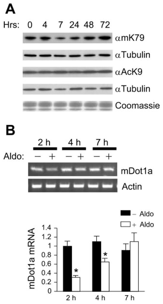
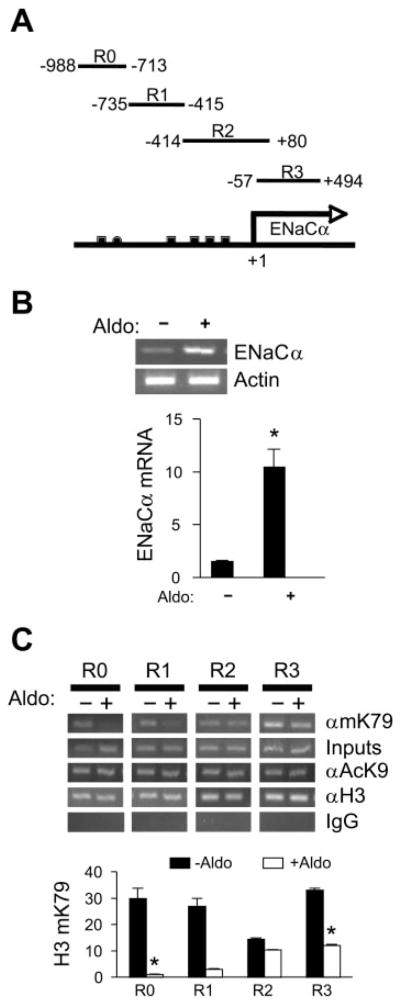
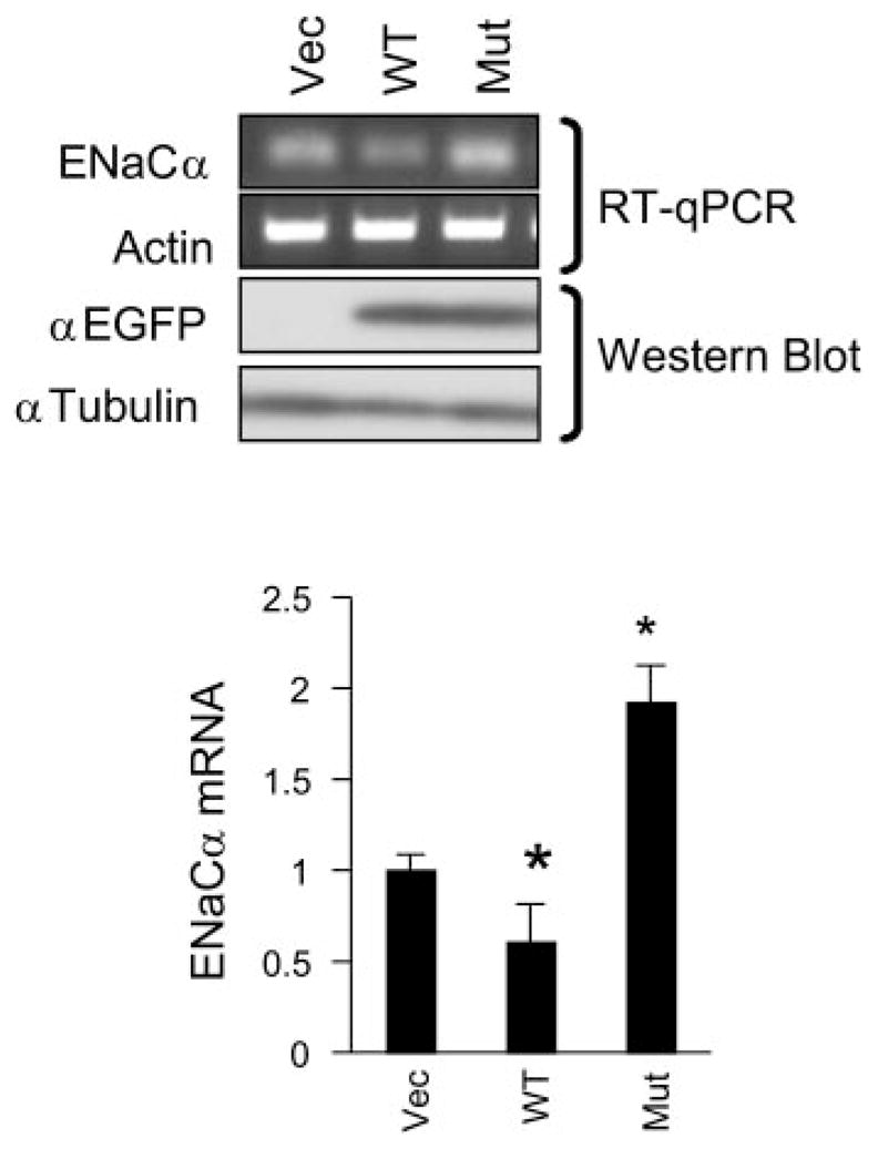
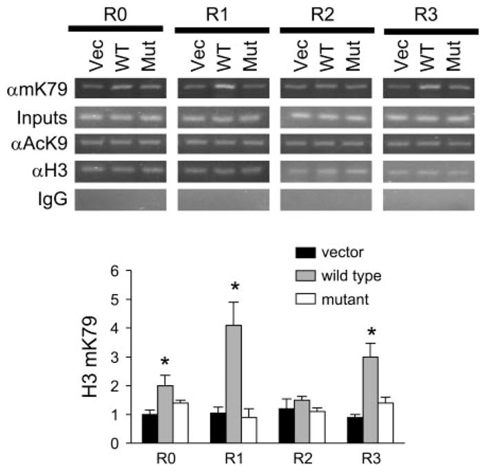
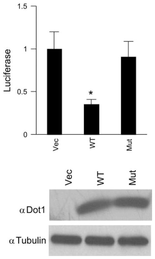
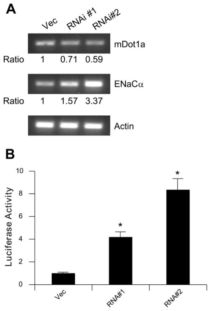
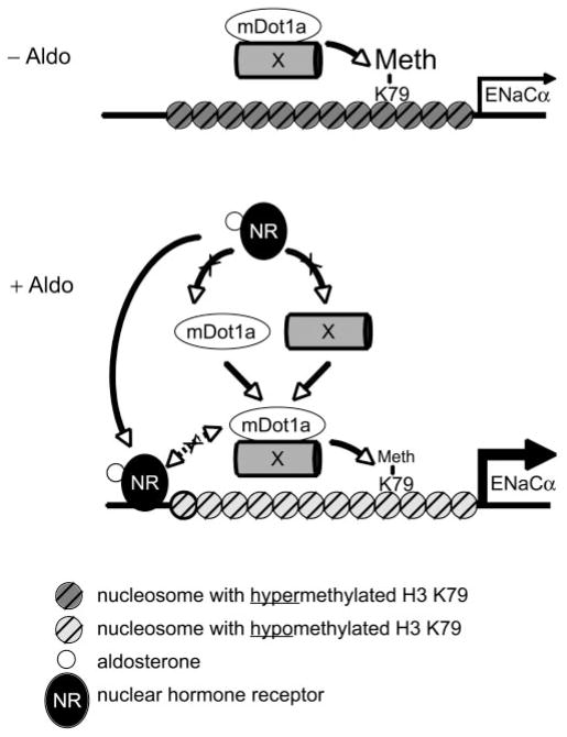
Aldosterone is a major regulator of epithelial Na(+) absorption. One of its principal targets is the epithelial Na(+) channel alpha-subunit (ENaCalpha), principally expressed in the kidney collecting duct, lung, and colon. Models of aldosterone-mediated trans-activation of the ENaCalpha gene have focused primarily on interactions of liganded nuclear receptors with the ENaCalpha gene promoter. Herein, we demonstrate that the murine histone H3 lysine-79 methyltransferase, murine disruptor of telomeric silencing alternative splice variant "a" (mDot1a), is a novel component in the aldosterone signaling network controlling transcription of the ENaCalpha gene. Aldosterone downregulated mDot1a mRNA levels in murine inner medullary collecting ducts cells, which was associated with histone H3 K79 hypomethylation in bulk histones and at specific sites in the ENaCalpha 5'-flanking region, and trans-activation of ENaCalpha. Knockdown of mDot1a by RNA interference increased activity of a stably integrated ENaCalpha promoter-luciferase construct and expression of endogenous ENaCalpha mRNA. Conversely, overexpression of EGFP-tagged mDot1a resulted in hypermethylation of histone H3 K79 at the endogenous ENaCalpha promoter, repression of endogenous ENaCalpha mRNA expression, and decreased activity of the ENaCalpha promoter-luciferase construct. mDot1a-mediated histone H3 K79 hypermethylation and repression of ENaCalpha promoter activity was abolished by mDot1a mutations that eliminate its methyltransferase activity. Collectively, our data identify mDot1a as a novel aldosterone-regulated histone modification enzyme, and, through binding the ENaCalpha promoter and hypermethylating histone H3 K79 associated with the ENaCalpha promoter, a negative regulator of ENaCalpha transcription.


Figures

Fig. 1
Fig. 1
Characterization of a murine disruptor of telomeric silencing alternative splice variant “a” (mDot1a)-specific antibody. Whole cell lysates from mouse kidney (lane 1) or from mouse inner meduallry collecting ducts (mIMCD3) cells transfected with either plasmid epidermal growth factor protein C3 (pEGFPC3) vector control (lanes 2, 4, 6, and 8) or pEGFPC3-mDot1a 2–478, encoding aa 2–478 of mDot1a fused to EGFP (lanes 3, 5, 7, and 9), were analyzed by Western blots with an antiserum against mDot1 (αDot1) or anti-EGFP antibody (αEGFP). As controls for specificity, blots were also prepared using αDot1 plus a molar excess of the immunizing peptide (Pep) or preimmune serum (Pre). Shorter exposures revealed that the band of ~170 kDa in the mouse kidney samples (lane 1) consists of a doublet (data not shown). The identity of the ~25 kDa protein that comigrated with EGFP (lane 8) and was weakly labeled in all cases is unknown and nonspecific (NS).
Fig. 2
Fig. 2
Aldosterone dynamically regulates the levels of Dot1a mRNA and H3 K79 methylation in mIMCD3 cells. A: acid extracts rich in histones isolated from mIMCD3 cells that had been cultured in charcoal-stripped serum and treated with 1 μM aldosterone for the indicated times were separated on identical gels, and immunoblotted with antibodies specific for methylated H3 at K79 (αmK79), acetylated H3 at K9 (αAcK9) or stained with Coomassie blue dye. The blots were stripped and reprobed with an antibody against α-tubulin (αTubulin). B: total RNA prepared from mIMCD3 cells treated with vehicle (ethanol) or 1 μM aldosterone for 2, 4, or 7 h was analyzed by agarose gel electrophoresis and by real-time RT-quantitative PCR (qPCR; presented in the histogram) with primers specific for mDot1a or actin. The mDot1a mRNA levels were normalized against invariant actin mRNA, as measured by real-time PCR. The mDot1a mRNA level of the vehicle-treated cells for 2 h was arbitrarily set as 1 and used to calculate the relative levels of all other samples. *P < 0.05 vs. −Aldo; n = 3.
Fig. 3
Fig. 3
Aldosterone induces hypomethylation of histone H3 K79 at the epithelial Na+ channel α-subunit (mENaCα) 5′-flanking region and ENaCα expression in mIMCD3 cells. mIMCD3 cells were cultured with charcoal-stripped serum and treated with either 1 μM aldosterone or vehicle for 2 h. Similar sets of cells were harvested for RT-qPCR, Western blot analysis, or chromatin immunoprecipitation (ChIP). A: diagram of the 5′-flanking region of mENaCα fragments designated R0-R3 are shown along with their relative positions to the major transcription start site (+1) of mENaCα. ● and ■ represent the putative GRE site (−811) and GRE half sites (−983, −416, −325, −241, and −234), respectively. B: the mENaCα mRNA levels were examined by agarose gel electrophoresis and RT-qPCR (see Fig. 2B). *P < 0.05 vs. −Aldo; n = 3. C: DNAs coimmunoprecipitated by the indicated antibodies were analyzed by agarose gel electrophoresis and by real-time qPCR (bottom histogram) with primers designed to specifically amplify R0 to R3, followed by agarose gel analysis. Antibodies against the acetylated histone H3 K9 (αAcK9), the NH2 terminal tail of histone H3 (αH3), and normal rabbit IgG were used as controls. The relative histone H3 K79 methylation was defined as ChIP efficiency, i.e., the immunoprecipitated amount of material to that of the input sample, as determined by real-time PCR and verified by agarose gel analysis. *P < 0.05 vs. −Aldo; n = 3.
Fig. 4
Fig. 4
Overexpression of wild-type mDot1a, but not its methyltransferase-dead mutant, decreases mENaCα mRNA levels in mIMCD3 cells. mIMCD3 cells were transiently transfected with an empty vector pEGFPC3 (Vec) or its derivatives expressing EGFP-tagged wild-type (WT) or methyltransferase-dead mutant (Mut). RNA samples were prepared and analyzed for ENaCα and actin expression by agarose gel electrophoresis and RT-qPCR (bottom histogram). Antibodies against EGFP (αEGFP) or tubulin (αTubulin) were used to monitor the expression of EGFP-mDot1a fusions and to verify equal loading on Western blots. *P < 0.05 vs. corresponding vector control; n = 3.
Fig. 5
Fig. 5
Overexpression of wild-type mDot1a, but not its methyltransferase-dead mutant, increases histone H3 K79 methylation at specific portions of the mENaCα 5′-flanking region in mIMCD3 cells. ChIP assays coupled with real-time qPCR were carried out with the indicated antibodies to determine the association of EGFP-mDot1a fusions with the R0-R3 subregions of the mENaCα 5′-flanking region. *P < 0.05 vs. corresponding vector control; n = 3.
Fig. 6
Fig. 6
Overexpressed mDot1a represses the activity of an mENaCα promoter-luciferase reporter gene in a methyltransferase-dependent manner. An mIMCD3-derived cell line harboring a stably transfected firefly luciferase reporter driven by the mENaCα promoter (−850 to +536, just before the translation start site ATG) was cultured in charcoal-stripped serum and transiently transfected with an empty vector pcDNA 3.1-V2 (Vec) or its derivatives expressing untagged wild-type mDot1a (WT) or methyltransferase-dead mDot1a mutant (Mut). Renilla luciferase reporter pRL-SV40 was included as an internal control. Firefly luciferase activity of each sample was normalized to its Renilla luciferase activity. The firefly luciferase activity of the vector-transfected cells was designated as 1 and utilized to determine the relative level and the significance of the other samples. *P < 0.05 vs. Vec; n = 3. Overexpressed mDot1a proteins were monitored by Western blot analyses with the αDot1 characterized in Fig. 1, and equal loading was verified by an identical blot probed with anti-tubulin.
Fig. 7
Fig. 7
Knockdown of mDot1a mRNA expression by RNA interference increases expression of endogenous mENaCα and a mENaCα promoter-luciferase reporter. A: three independent cell lines stably transfected with pSilencer4.1-CMV-neo negative control (Vec) or mDot1a silencing constructs pSCN-RNAi1 (RNAi#1) or pSCN-RNAi2 (RNAi#2) were examined by real-time RT-qPCR for expression of mDot1a, mENaCα, or actin. The level of mDot1a and mENaCα was first normalized to that of the actin in the same sample and then compared with that of the control cells. B: the same cells as in A were transiently transfected with the same mENaCα promoter-luciferase reporter, followed by luciferase assay as in Fig 6.
Fig. 8
Fig. 8
Histone H3 methylation at K79 occurs in nuclei of kidney collecting duct cells. Tissue sections of the mouse kidney were fixed and indirect immunofluorescence performed as detailed in MATERIALS AND METHODS using rabbit polyclonal histone H3 dimethylK79 antiserum and anti-aquaporin-2 raised in chickens as primary antibodies, with Alexa Fluor 488 goat anti-rabbit IgG and Alexa Fluor 594 goat anti-chicken IgG as secondary antibodies. Nuclei were stained with 4′,6-diamidino-2-phenylindole (DAPI). Samples without primary antibodies were used as a negative control (Neg. Control). Image analysis was performed as detailed in the MATERIALS AND METHODS. A representative image of the renal outer medulla is shown (magnification ×600); n = 3.
Fig. 9
Fig. 9
Hypothetical model for aldosterone-sensitive repression of ENaCα expression by an mDot1a-containing complex modulating histone H3 K79 hypermethylation associated with the ENaCα promoter. Under basal conditions, mDot1a and its putative interacting partner “X” form a nuclear complex that directly or indirectly binds to the 5′-flanking region of ENaCα, leading to hypermethylation of histone H3 K79 at this location and repression of ENaCα (A). In this model, aldosterone (○) stimulates ENaCα transcription by regulating two antagonistic complexes. Aldosterone binds and activates the nuclear hormone receptors (NR) that are either glucocorticoid receptor and/or miner-alocorticoid receptor (MR) homo- or heterodimers to bind the GRE for transactivation of ENaCα. In parallel, aldosterone downregulates the amount of the Dot1a complex, possibly by multiple mechanisms, including reducing the abundance of mDot1a and its partner X (shown) via NR-dependent (shown) or NR-independent mechanisms (not shown) to limit the mDot1a complex availability, leading to histone H3 K79 hypomethylation at specific ENaCα promoter subregions and release of repression of ENaCα. In either case, free mDot1a binds DNA nonspecifically and catalyzes histone H3 K79 methylation throughout the genome at the basal level (not shown). The putative counter-interaction between the NR-aldosterone complex and the mDot1a-“X” complex is shown with a dotted line and may tune the ultimate level of ENaCα transcription. The relative position of the site(s) associated with the mDot1a complex to the ENaCα transcription start site and GRE remains to be determined. Similar mechanisms may be applicable to other aldosterone-regulated genes.

Similar articles

Cited by

References

    1. Alvarez de la Rosa D, Li H, Canessa CM. Effects of aldosterone on biosynthesis, traffic, and functional expression of epithelial sodium channels in A6 cells. J Gen Physiol. 2002;119:427–442. - PMC - PubMed
    1. Asher C, Garty H. Aldosterone increases the apical Na+ permeability of toad bladder by two different mechanisms. Proc Natl Acad Sci USA. 1988;85:7413–7417. - PMC - PubMed
    1. Asher C, Wald H, Rossier BC, Garty H. Aldosterone-induced increase in the abundance of Na+ channel subunits. Am J Physiol Cell Physiol. 1996;271:C605–C611. - PubMed
    1. Boyd C, Naray-Fejes-Toth A. Gene regulation of ENaC subunits by serum- and glucocorticoid-inducible kinase-1. Am J Physiol Renal Physiol. 2005;288:F505–F512. - PubMed
    1. Cheung P, Tanner KG, Cheung WL, Sassone-Corsi P, Denu JM, Allis CD. Synergistic coupling of histone H3 phosphorylation and acetylation in response to epidermal growth factor stimulation. Mol Cell. 2000;5:905–915. - PubMed

Publication types

LinkOut - more resources