. 2005 Nov;187(21):7193-203.
doi: 10.1128/JB.187.21.7193-7203.2005.

Competition and coexistence between Streptococcus mutans and Streptococcus sanguinis in the dental biofilm

Affiliations

Competition and coexistence between Streptococcus mutans and Streptococcus sanguinis in the dental biofilm

Jens Kreth et al. J Bacteriol. 2005 Nov.

Abstract

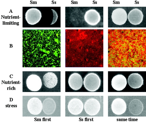
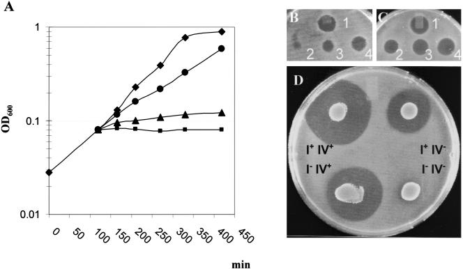
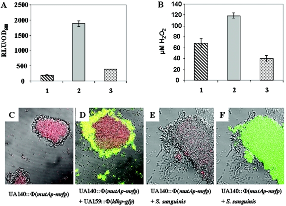
The human mucosal surface is colonized by the indigenous microflora, which normally maintains an ecological balance among different species. Certain environmental or biological factors, however, may trigger disruption of this balance, leading to microbial diseases. In this study, we used two oral bacterial species, Streptococcus mutans and Streptococcus sanguinis (formerly S. sanguis), as a model to probe the possible mechanisms of competition/coexistence between different species which occupy the same ecological niche. We show that the two species engage in a multitude of antagonistic interactions temporally and spatially; occupation of a niche by one species precludes colonization by the other, while simultaneous colonization by both species results in coexistence. Environmental conditions, such as cell density, nutritional availability, and pH, play important roles in determining the outcome of these interactions. Genetic and biochemical analyses reveal that these interspecies interactions are possibly mediated through a well-regulated production of chemicals, such as bacteriocins (produced by S. mutans) and hydrogen peroxide (produced by S. sanguinis). Consistent with the phenotypic characteristics, production of bacteriocins and H2O2 are regulated by environmental conditions, as well as by juxtaposition of the two species. These sophisticated interspecies interactions could play an essential part in balancing competition/coexistence within multispecies microbial communities.


Figures

FIG. 1.
FIG. 1.
Inhibition of oral streptococcal species by S. mutans UA140. 1, S. gordonii; 2, S. pyogenes; 3, S. oralis; 4, S. mitis ATCC 33399; 5, S. mitis ATCC 903; 6, S. pneumoniae; 7, S. cristatus; 8, S. parasanguinis; 9, S. sanguinis ATCC 10556; 10, S. sanguinis NY101; 11, S. sobrinus.
FIG. 2.
FIG. 2.
Competition assays between S. mutans and S. sanguinis. (A) Competition assay on half-strength BHI plate. (B) Confocal laser scanning microscopy analysis of competition in biofilms. Green cells, S. mutans (green fluorescent protein); red cells, S. sanguinis (Cell-tracker orange). The pictures were taken at ×100 magnification. (C) competition assays on “nutrient-rich” plate (BHI plus 1% sucrose, buffered to pH 7). (D) competition assays on “stress” plate (BHI at pH 5.5). (C and D) Left, S. mutans (Sm) was inoculated first; middle, S. sanguinis (Ss) was inoculated first; right Sm and Ss were inoculated at the same time.
FIG. 3.
FIG. 3.
Identification of inhibitory substances produced by S. mutans and S. sanguinis. (A) S. sanguinis (Ss) was inoculated first. (B) S. mutans (Sm) was inoculated first. After 24-h growth on half-strength BHI plates, 40 μg of peroxidase (left), 64 μg of peptidase (middle), or phosphate-buffered saline (right) was added beside the colony before the competing species was inoculated. (C) competition of the mutacin-defective strain UA140IIV with Ss on the plate (C) and in the biofilm (D) when Sm was inoculated first. Green cells, S. mutans (green fluorescent protein); red cells, S. sanguinis (Cell-tracker orange). The confocal micrograph was taken at ×100 magnification.
FIG. 4.
FIG. 4.
Effects of H2O2, mutacin I, and mutacin IV on the growth of S. mutans and S. sanguinis. (A) Growth inhibition of S. mutans UA140 treated with different concentrations of H2O2; ⧫, no H2O2; •, 0.0005% (142 μM) H2O2; ▴, 0.0025% (710 μM) H2O2; ▪, 0.005% (1.42 mM) H2O2. Experiments were repeated two times with similar results. Shown is a representative result of one experiment. (B and C) Inhibition of S. sanguinis with purified mutacin I and partially purified mutacin IV. Different dilutions of purified mutacin I (B) and partially purified mutacin IV (C) were spotted onto a BHI plate and overlaid with S. sanguinis. Each spot contained 10 μl of twofold serially diluted extract (i.e., no. 1, undiluted; no. 4, twofold diluted; no. 3, fourfold diluted, etc.). (D) Effects of mutations in mutacin I and mutacin IV genes on the growth of S. sanguinis. Overnight cultures of a mutacin I-defective (IIV+), a mutacin IV-defective (I+IV), a double-mutant (IIV), and a wild-type (I+IV+) strain of UA140 were spotted (10 μl) onto BHI plates and overlaid with S. sanguinis.
FIG. 5.
FIG. 5.
Effects of growth conditions on mutacin I gene expression (A), mutacin production (B), and H2O2 production (C). Mutacin I gene expression (mutAp-luc) was measured as relative light units (RLU) per OD600 unit; mutacin production was measured by diameters of the inhibition zone against the indicator; H2O2 production by S. sanguinis was indicated by a purple color (see Materials and Methods). Cells were grown on different conditioned plates: 1, half-strength BHI; 2, BHI plus 1% sucrose, pH 7; 3, BHI, pH 5.5. Presented are representatives of at least two experiments performed on different days (the error bars indicate standard deviations).
FIG. 6.
FIG. 6.
Effects of juxtaposition between S. mutans and S. sanguinis on mutacin gene expression and H2O2 production. (A) Mutacin I gene expression of strain UA140::Φ(mutAp-luc). (B) H2O2 production by S. sanguinis. Bars 1, single-species planktonic culture; bars 2, single-species pelleted culture; bars 3, mixed-species pelleted culture. The experiment was done three times on different days with similar results. Presented are representative data from one experiment done in duplicate. The error bars indicate standard deviations. (C) Mutacin I gene expression in strain UA140::Φ(mutAp-mrfp) single-strain culture. Red fluorescence indicates mutacin I gene expression. (D) Same experiment as in panel C, but mixed with strain UA159::Φ(ldhp-gfp). Red, UA140::Φ(mutAp-mrfp); green, UA159::Φ(ldhp-gfp); yellow, mixture of the two strains. (E) Same experiment as in panel C, but mixed with S. sanguinis. (F) Same experiment as in panel E with UA140::Φ(mutAp-mrfp) cells labeled with fluorescein isothiocyanate-conjugated anti-S. mutans monoclonal antibodies. Green cells, UA140::Φ(mutAp-mrfp); gray cells, S. sanguinis. The confocal micrographs were taken with fluorescent and differential interference contrast modes.
FIG. 7.
FIG. 7.
Relative mutacin I (mutAp-luc) and lactate-dehydrogense (ldhp-luc) gene expression in single- and mixed-species surface biofilms. Overnight cultures of all strains were adjusted to an OD600 of 1. Ten microliters of strain UA140::Φ(mutAp-luc) or UA140::Φ(ldhp-luc) alone (bars 1 and 3) or mixed in a 1:1 ratio with S. sanguinis (bars 2 and 4) were spotted onto a BHI plate. After 6 h of incubation, the cells were scraped from the plate and the luciferase activity was determined. The activity was normalized by the cell counts of S. mutans after serial dilution. Experiments were repeated twice with similar results. Shown is a representative result of one experiment done with triplicate samples. (A) Expression of the mutacin I (mutA) gene. (B) Expression of the ldh gene.

Similar articles

Cited by

References

    1. Ajdic, D., W. M. McShan, R. E. McLaughlin, G. Savic, J. Chang, M. B. Carson, C. Primeaux, R. Tian, S. Kenton, H. Jia, S. Lin, Y. Qian, S. Li, H. Zhu, F. Najar, H. Lai, J. White, B. A. Roe, and J. J. Ferretti. 2002. Genome sequence of Streptococcus mutans UA159, a cariogenic dental pathogen. Proc. Natl. Acad. Sci. USA 99:14434-14439. - PMC - PubMed
    1. Becker, M. R., B. J. Paster, E. J. Leys, M. L. Moeschberger, S. G. Kenyon, J. L. Galvin, S. K. Boches, F. E. Dewhirst, and A. L. Griffen. 2002. Molecular analysis of bacterial species associated with childhood caries. J. Clin. Microbiol. 40:1001-1009. - PMC - PubMed
    1. Bender, G. R., S. V. Sutton, and R. E. Marquis. 1986. Acid tolerance, proton permeabilities, and membrane ATPases of oral streptococci. Infect. Immun. 53:331-338. - PMC - PubMed
    1. Blehert, D. S., R. J. Palmer, Jr., J. B. Xavier, J. S. Almeida, and P. E. Kolenbrander. 2003. Autoinducer 2 production by Streptococcus gordonii DL1 and the biofilm phenotype of a luxS mutant are influenced by nutritional conditions. J. Bacteriol. 185:4851-4860. - PMC - PubMed
    1. Caufield, P. W., A. P. Dasanayake, Y. Li, Y. Pan, J. Hsu, and J. M. Hardin. 2000. Natural history of Streptococcus sanguinis in the oral cavity of infants: evidence for a discrete window of infectivity. Infect. Immun. 68:4018-4023. - PMC - PubMed

Publication types

MeSH terms

LinkOut - more resources