. 2005 Nov 1;175(9):5799-808.
doi: 10.4049/jimmunol.175.9.5799.

Recognition of fresh human tumor by human peripheral blood lymphocytes transduced with a bicistronic retroviral vector encoding a murine anti-p53 TCR

Affiliations

Recognition of fresh human tumor by human peripheral blood lymphocytes transduced with a bicistronic retroviral vector encoding a murine anti-p53 TCR

Cyrille J Cohen et al. J Immunol. .

Erratum in

  • J Immunol. 2006 Oct 15;177(8):5746

Abstract

The p53 protein is markedly up-regulated in a high proportion of human malignancies. Using an HLA-A2 transgenic mouse model, it was possible to isolate high-avidity murine CTLs that recognize class I-restricted human p53 epitopes. We isolated the alpha- and beta-chain of a TCR from a highly avid murine CTL clone that recognized the human p53(264-272) epitope. These genes were cloned into a retroviral vector that mediated high efficiency gene transfer into primary human lymphocytes. Efficiencies of >90% for gene transfer into lymphocytes were obtained without selection for transduced cells. The p53 TCR-transduced lymphocytes were able to specifically recognize with high-avidity, peptide-pulsed APCs as well as HLA-A2.1+ cells transfected with either wild-type or mutant p53 protein. p53 TCR-transduced cells demonstrated recognition and killing of a broad spectrum of human tumor cell lines as well as recognition of fresh human tumor cells. Interestingly, both CD8+ and CD4+ subsets were capable of recognizing and killing target cells, stressing the potential application of such a CD8-independent TCR molecule that can mediate both helper and cytotoxic responses. These results suggest that lymphocytes genetically engineered to express anti-p53 TCR may be of value for the adoptive immunotherapy of patients with a variety of common malignancies.


Conflict of interest statement

Disclosures

The authors have no financial conflict of interest.

Figures

FIGURE 1
FIGURE 1
Functional analysis of p53 TCR. A, RNA electroporation of stimulated PBL. PBLs stimulated with OKT3 Ab plus IL-2 were electroporated with in vitro-transcribed RNA at 2 μg/1 × 106 cells. Twenty hours after electroporation, murine TCR expression was determined by FACS analysis for PBLs electroporated with RNA encoding p53 TCR that were stained with anti-murine TCR (1), with isotype control (2), and for non-electroporated cells stained with anti-murine TCR (3). B, PBLs were elec-troporated with p53 TCR RNA and cocultured with peptide-pulsed T2 cells. Peptides used were HLA-A2-restricted p53264 –272 and NY-ESO-1157–165V. C, Schematic representation of the retroviral vector encoding the p53 TCR. LTR, Long terminal repeat.
FIGURE 2
FIGURE 2
Efficiency of the retroviral transduction. Human PBLs were transduced with the retroviral vector encoding the p53 TCR. A–D, Expression was assessed by FACS 48 h after transduction. The percentage of PBLs that express the mouse TCR (mTCR) of interest are as shown.
FIGURE 3
FIGURE 3
Recognition of peptide-pulsed and p53-transfected cells. A, Human PBLs expressing the p53 TCR were cocultured for 16 h with T2 cells pulsed with different concentrations of p53-specific peptide (p53264–272). The concentration of IFN-γ secreted in the medium was measured using an ELISA procedure. IFN-γ secretion in cultures with T2 pulsed with control peptides (gp100–209, gp100–280, MART-126–35, p53149–157, and HBVc) was ≤480 pg/ml (data not shown). B, Human PBLs expressing the p53 TCR were cocultured for 16 h with CosA2 cells that were transfected with different p53 (both wt and mutant) plasmid expression vectors. The concentration of IFN-γ secreted in the medium was measured using ELISA.
FIGURE 4
FIGURE 4
Recognition of human tumor lines. Human PBLs expressing the p53 TCR were cocultured for 16 h with the indicated tumor cell lines. The concentration of IFN-γ secreted in the medium was measured using an ELISA.
FIGURE 5
FIGURE 5
Tumor cell-mediated IL-2 synthesis. Human PBLs expressing the p53 TCR were cocultured for 16 h with the indicated tumor cell lines. The concentration of IL-2 secreted in the medium was measured using an ELISA.
FIGURE 6
FIGURE 6
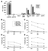
In vitro proliferation of TCR-engineered PBLs. Human PBLs expressing the p53 TCR were labeled with CFSE and cocultured with tumor cell lines. Ag-specific proliferation was measured after 3 (A and C) and 6 (B and D) days as shown by decreasing CFSE fluorescence of mock-transduced (A and B) or p53 TCR-expressing (C and D) PBLs cocultured with Saos 2, H2087, MDA-MB-231, or no target.
FIGURE 7
FIGURE 7
Functional reactivity of p53 TCR-transduced lymphocytes. Expression of CD107a (degranulation marker) was detected by FACS analysis on the surface of human PBLs expressing the p53 TCR after 2 h coculture with the indicated tumor cell line (control: A–D; p53+/HLA-A2+ : E–K). The percentage of p53 TCR+/CD107a+ cells was as shown.
FIGURE 8
FIGURE 8
Specific killing of tumor cell lines. Human PBLs expressing the p53 TCR were cocultured for 6 h with the indicated tumor cell lines previously labeled with 51Cr. Specific lysis was measured at the E:T ratio indicated using: [(specific release − spontaneous release)/(total release − spontaneous release)]. A, The HLA-A2+/p53+ cell lines used include: Saos 2/*143 (▴), MDA-MB-231 (×), H2087 (♦), BE-3 (•), in addition to the two controls MDA 386 (*, HLA-A2/p53+) and Saos 2 (▪, HLA-A2+/p53). B, We compared the lysis activity of p53 TCR-transduced cells (filled symbols) with mock-transduced cells (open symbols) on the following targets: HLA-A2+/p53+ H2087 (diamond), Saos 2 (square), normal renal cells ELW91 (triangle), OKT3/IL-2 activated PBLs (circle), and normal resting (3 wk poststimulation) (ellipse) T cells.
FIGURE 9
FIGURE 9
Specific recognition of fresh human tumor cells. Human PBLs expressing the p53 TCR were cocultured for 16 h with fresh human tumor cells (melanoma) from different patients. The concentration of IFN-γ secreted in the medium was measured using an ELISA procedure.
FIGURE 10
FIGURE 10
Recognition and killing of tumor by transduced CD4+ cells. A, Different subsets (CD8+, CD4+, or bulk population) of human PBLs expressing the p53 TCR were cocultured for 16 h with the indicated peptide-pulsed APCs, tumor cell lines, or fresh human tumor samples (patient no. 7, HLA-A2+ and patient no. 12, HLA-A2). The concentration of IFN-γ secreted in the medium was measured by ELISA. B–E, Different T cell subsets (CD8+, CD4+, or bulk population) of human PBLs expressing the p53 TCR were cocultured for 6 h with different tumor cell lines previously labeled with 51Cr. Specific lysis of Saos 2 (B), H2087 (C), MDA-MB-231 (D), JY (E) was measured at the E:T ratio indicated using: [(specific release − spontaneous release)/(total release − spontaneous release)].

References

    1. Berzofsky, J. A., L. J. Helman, and D. P. Carbone. 2000. Cancer vaccines: cancer antigens, oncogenes and mutations. In Principles and Practices of the Biologic Therapy of Cancer. S. A. Rosenberg, ed. Lippincott Williams & Wilkins, Philadelphia, pp. 526–541.
    1. Levine AJ, Momand J, Finlay CA. The p53 tumour suppressor gene. Nature. 1991;351:453–456. - PubMed
    1. Olive KP, Tuveson DA, Ruhe ZC, Yin B, Willis NA, Bronson RT, Crowley D, Jacks T. Mutant p53 gain of function in two mouse models of Li-Fraumeni syndrome. Cell. 2004;119:847–860. - PubMed
    1. Lang GA, Iwakuma T, Suh YA, Liu G, Rao VA, Parant JM, Valentin-Vega YA, Terzian T, Caldwell LC, Strong LC, et al. Gain of function of a p53 hot spot mutation in a mouse model of Li-Fraumeni syndrome. Cell. 2004;119:861–872. - PubMed
    1. Winter SF, Minna JD, Johnson BE, Takahashi T, Gazdar AF, Carbone DP. Development of antibodies against p53 in lung cancer patients appears to be dependent on the type of p53 mutation. Cancer Res. 1992;52:4168–4174. - PubMed

Substances