

doi: 10.1093/nar/gni150.
Proximity ligation assays with peptide conjugate 'burrs' for the sensitive detection of spores
Affiliations
- PMID: 16237122
- PMCID: PMC1258177
- DOI: 10.1093/nar/gni150
Item in Clipboard
Proximity ligation assays with peptide conjugate 'burrs' for the sensitive detection of spores
Nucleic Acids Res.
.
Abstract
The proximity ligation assay (PLA) has previously been used for the sensitive and specific detection of single proteins. In order to adapt PLA methods for the detection of cell surfaces, we have generated multivalent peptide-oligonucleotide-phycoerythrin conjugates ('burrs') that can bind adjacent to one another on a cell surface and be ligated together to form unique amplicons. Real-time PCR detection of burr ligation events specifically identified as few as 100 Bacillus anthracis, 10 Bacillus subtilis and 1 Bacillus cereus spore. Burrs should prove to be generally useful for detecting and mapping interactions and distances between cell surface proteins.
Figures

Specificity of monovalent and polyvalent probes. Fluorescent probes were constructed using the NH-peptide (BS-specific). BS and BC spores were incubated with either (A) monovalent NH-peptide–fluorescein conjugates or (B) polyvalent NH-peptide–PE conjugates. Specific binding was only observed when the polyvalent NH-peptide–PE probes were used. Spores were visualized using differential interference microscopy (DIC) and fluorescence microscopy with either fluorescein (FITC) or Texas Red filter sets (TR).

Construction and ligation of burrs. (A) Burrs: oligonucleotides and peptides are separately conjugated to PE. There are two distinct oligonucleotide conjugates, one linked through its 5′ end and one linked through its 3′ end. (B) Burr ligation and amplification. When simultaneously bound to a spore target, burrs can be aligned by a splint oligonucleotide and ligated to generate a unique amplicon. The sequences of the 5′ and 3′ oligonucleotide probes as well as the splint oligonucleotide are given in Materials and Methods.

Optimization of PLA probe concentration for the detection of B.cereus spores. The real-time PCR data represent a single dataset in which the probe concentration was varied from 0.1 to 100 pM. PLA reactions conducted in the presence of 100 BC spores are indicated by a solid line and those conducted in the absence of spores by dashed lines. A positive, spore-dependent signal was only observed when reactions were conducted using 10 pM probe (bold red). Reactions were assembled as described in Materials and Methods.

Optimization of PLA probe concentration for 100 B.subtilis and B.cereus spores. A splint concentration of 10 pM was used. The cycle difference represents the difference between the C[T] value of the background amplification reaction (no spores) and amplification in the presence of spores. Reactions containing BS spores and BC spores were carried out with burrs that presented either the NH- (BS-specific) or S-peptides (BC-specific). Reactions containing BA (Sterne) spores were carried out with burrs presenting either the NH-, S- or the ATY-peptides (BA-specific).

Splint optimization for 100 spores. A probe concentration of 1 pM was used. Reactions contained burrs as described in Figure 4. Cycle difference is as in Figure 4.
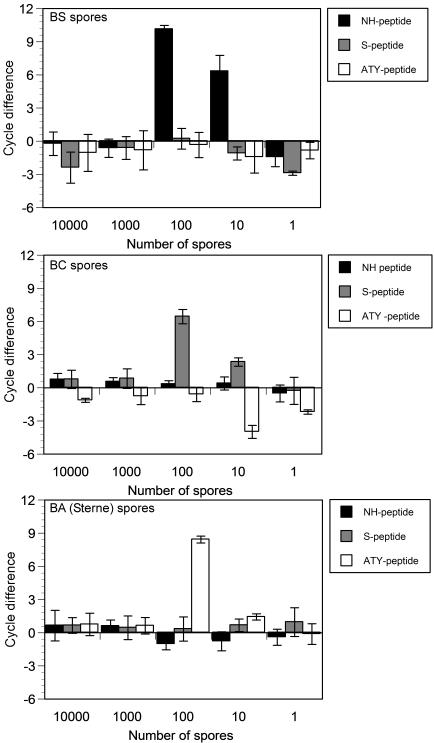
Specificity of spore detection assays. Reactions were carried out with 10 pM probe and 10 pM splint, and contained burrs bearing one of the three spore-specific peptides. Cycle difference is as in Figure 4.

Optimization of the limits of detection for BC spores. Reactions were carried out with 10 pM probe and 1 pM splint, and contained burrs bearing one of the three spore-specific peptides. Cycle difference is as in Figure 4.
Similar articles
-
Peptides panned from a phage-displayed random peptide library are useful for the detection of Bacillus anthracis surrogates B. cereus 4342 and B. anthracis Sterne.Biochem Biophys Res Commun. 2010 Apr 23;395(1):93-8. doi: 10.1016/j.bbrc.2010.03.145. Epub 2010 Mar 27. Biochem Biophys Res Commun. 2010. PMID: 20350526
-
Spore-forming organisms in platelet concentrates: a challenge in transfusion bacterial safety.Transfus Med. 2008 Dec;18(6):371-6. doi: 10.1111/j.1365-3148.2008.00895.x. Transfus Med. 2008. PMID: 19140821
-
Evaluation of different methods to discriminate Bacillus anthracis from other bacteria of the Bacillus cereus group.J Appl Microbiol. 2006 Apr;100(4):673-81. doi: 10.1111/j.1365-2672.2006.02809.x. J Appl Microbiol. 2006. PMID: 16553722
-
Discovery of phage display peptide ligands for species-specific detection of Bacillus spores.J Microbiol Methods. 2003 May;53(2):263-71. doi: 10.1016/s0167-7012(03)00030-7. J Microbiol Methods. 2003. PMID: 12654497 Review.
-
Spores of Bacillus subtilis: their resistance to and killing by radiation, heat and chemicals.J Appl Microbiol. 2006 Sep;101(3):514-25. doi: 10.1111/j.1365-2672.2005.02736.x. J Appl Microbiol. 2006. PMID: 16907802 Review.
Cited by
-
Phage display--a powerful technique for immunotherapy: 2. Vaccine delivery.Hum Vaccin Immunother. 2012 Dec 1;8(12):1829-35. doi: 10.4161/hv.21704. Epub 2012 Aug 21. Hum Vaccin Immunother. 2012. PMID: 22906938 Free PMC article. Review.
-
Deoxyribozymes that recode sequence information.Nucleic Acids Res. 2006 Apr 28;34(8):2166-72. doi: 10.1093/nar/gkl176. Print 2006. Nucleic Acids Res. 2006. PMID: 16648360 Free PMC article.
-
Combining Deep Phenotyping of Serum Proteomics and Clinical Data via Machine Learning for COVID-19 Biomarker Discovery.Int J Mol Sci. 2022 Aug 15;23(16):9161. doi: 10.3390/ijms23169161. Int J Mol Sci. 2022. PMID: 36012423 Free PMC article.
-
Streamlined circular proximity ligation assay provides high stringency and compatibility with low-affinity antibodies.Proc Natl Acad Sci U S A. 2018 Jan 30;115(5):E925-E933. doi: 10.1073/pnas.1718283115. Epub 2018 Jan 16. Proc Natl Acad Sci U S A. 2018. PMID: 29339495 Free PMC article.
-
DNAzyme-Functionalized R-Phycoerythrin as a Cost-Effective and Environment-Friendly Fluorescent Biosensor for Aqueous Pb2+ Detection.Sensors (Basel). 2019 Jun 18;19(12):2732. doi: 10.3390/s19122732. Sensors (Basel). 2019. PMID: 31216658 Free PMC article.
References
-
- Saiki R.K., Scharf S., Faloona F., Mullis K.B., Horn G.T., Erlich H.A., Arnheim N. Enzymatic amplification of beta-globin genomic sequences and restriction site analysis for diagnosis of sickle cell anemia. Science. 1985;230:1350–1354. - PubMed
-
- Ellerbrok H., Nattermann H., Ozel M., Beutin L., Appel B., Pauli G. Rapid and sensitive identification of pathogenic and apathogenic Bacillus anthracis by real-time PCR. FEMS Microbiol. Lett. 2002;214:51–59. - PubMed
-
- Ramisse V., Patra G., Garrigue H., Guesdon J.L., Mock M. Identification and characterization of Bacillus anthracis by multiplex PCR analysis of sequences on plasmids pXO1 and pXO2 and chromosomal DNA. FEMS Microbiol. Lett. 1996;145:9–16. - PubMed
-
- Ryu C., Lee K., Yoo C., Seong W.K., Oh H.B. Sensitive and rapid quantitative detection of anthrax spores isolated from soil samples by real-time PCR. Microbiol. Immunol. 2003;47:693–699. - PubMed
-
- Beyer W., Pocivalsek S., Bohm R. Polymerase chain reaction–ELISA to detect Bacillus anthracis from soil samples—limitations of present published primers. J. Appl. Microbiol. 1999;87:229–236. - PubMed
Publication types
MeSH terms
Substances
LinkOut - more resources
Full Text Sources
Other Literature Sources
Molecular Biology Databases
