

Transplantation of neuronal and glial restricted precursors into contused spinal cord improves bladder and motor functions, decreases thermal hypersensitivity, and modifies intraspinal circuitry
- PMID: 16237167
- PMCID: PMC6725721
- DOI: 10.1523/JNEUROSCI.2175-05.2005
Transplantation of neuronal and glial restricted precursors into contused spinal cord improves bladder and motor functions, decreases thermal hypersensitivity, and modifies intraspinal circuitry
Abstract
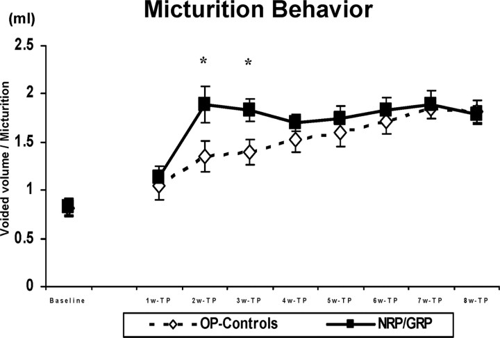
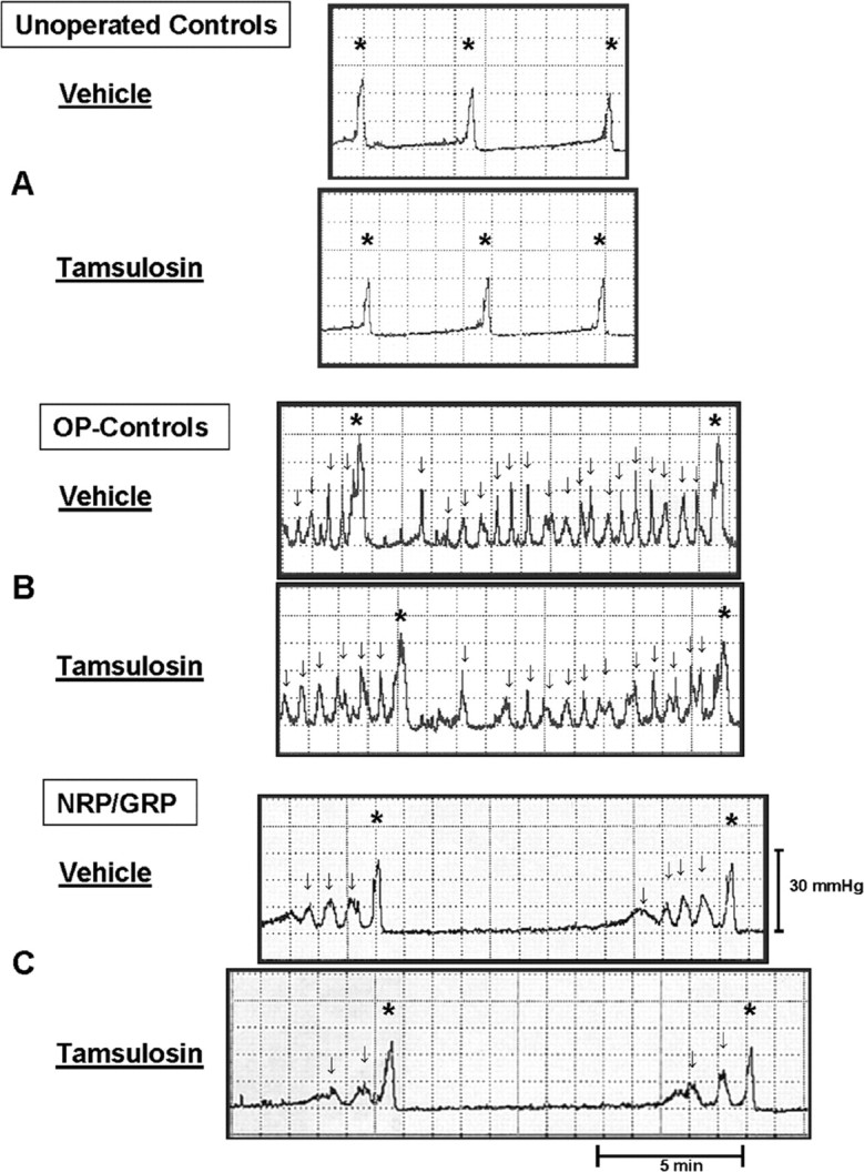
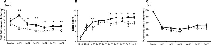
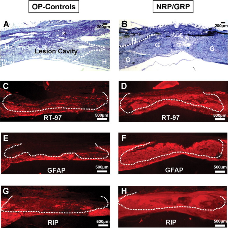
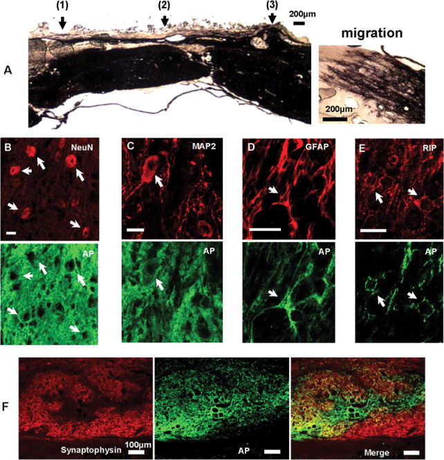
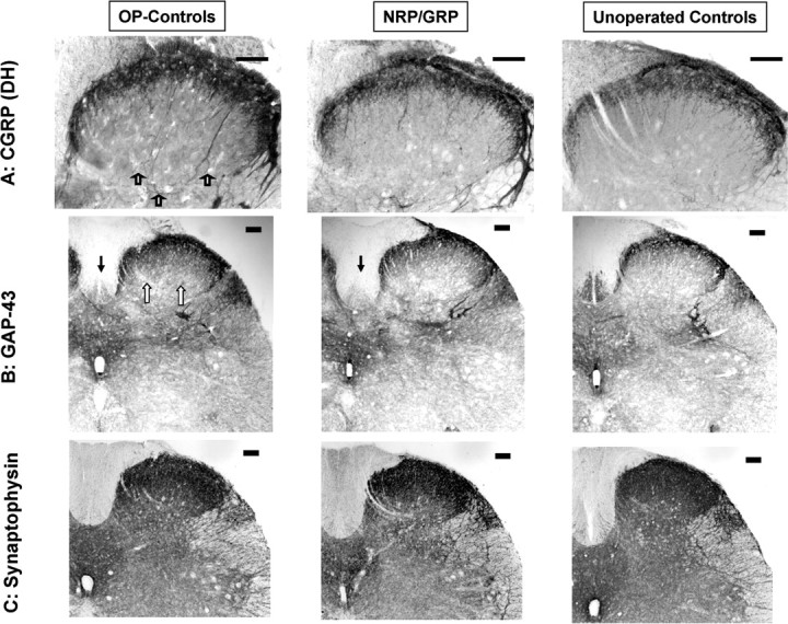
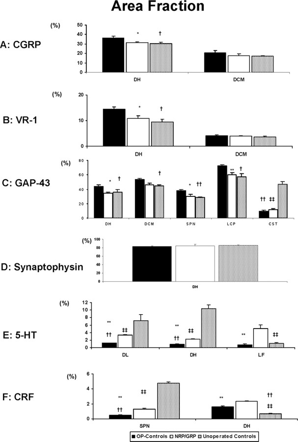
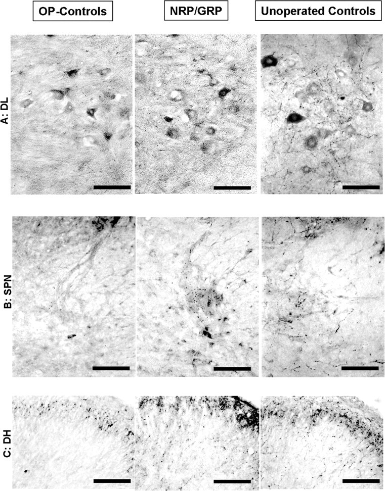
Transplanting neuronal and glial restricted precursors (NRP/GRP) into a midthoracic injury 9 d after contusion improved bladder and motor function, diminished thermal hypersensitivity, and modified lumbosacral circuitry compared with operated controls (OP-controls). Histological analysis showed that NRP/GRP survived, filled the lesion site, differentiated into neurons and glia, and migrated selectively. Volume of spinal cord spared was increased in NRP/GRP recipients, suggesting local protection. Bladder areflexia developed in both operated groups, but NRP/GRP recipients exhibited an accelerated recovery, with decreased micturition pressure and fewer episodes of detrusor hyperreflexia. Because noradrenergic receptors proliferate after spinal injury and descending noradrenergic pathways contribute to regulation of bladder control, we examined the effects of administering an alpha-1A-adrenergic antagonist, Tamsulosin, on urodynamics. This improved all cystometric parameters in both operated groups, and micturition pressure in NRP/GRP rats recovered to normal levels. Both operated groups initially showed increased sensitivity to a thermal stimulus applied to the tail; the NRP/GRP rats showed significant improvement over time. NRP/GRP grafts also produced greater recovery of hindlimb function in several tests, although both groups showed persistent and similar deficits in locomotion on a grid. Because bladder, hindlimb, and tail sensory and motor functions are organized through lumbosacral cord, we examined descending and primary afferent projections at L6-S1. The density of serotonergic, noradrenergic, and corticotrophin releasing factor-positive fibers increased in the NRP/GRP group compared with OP-controls, suggesting some sparing and/or sprouting of these modulatory pathways. Immunocytochemical staining density of dorsal root axons in the dorsal horn increased in the OP-controls but appeared normal in the NRP/GRP group. Synaptophysin immunoreactivity in the lumbosacral dorsal horn was similar among groups, consistent with restoration of synaptic density in both groups of operated animals but by different pathways. We suggest that local protection provided by NRP/GRP resulted in increased sparing/sprouting of descending pathways, which prevented sprouting by dorsal root axons, and that this modification in lumbosacral circuitry contributes to the recovery of function.
Figures
Similar articles
-
Acute administration of AMPA/Kainate blocker combined with delayed transplantation of neural precursors improves lower urinary tract function in spinal injured rats.Brain Res. 2011 Oct 18;1418:23-31. doi: 10.1016/j.brainres.2011.08.023. Epub 2011 Aug 22. Brain Res. 2011. PMID: 21937028 Free PMC article.
-
Transplants of fibroblasts expressing BDNF and NT-3 promote recovery of bladder and hindlimb function following spinal contusion injury in rats.Exp Neurol. 2005 Aug;194(2):410-31. doi: 10.1016/j.expneurol.2005.02.022. Exp Neurol. 2005. PMID: 16022868
-
Glial restricted precursor cell transplant with cyclic adenosine monophosphate improved some autonomic functions but resulted in a reduced graft size after spinal cord contusion injury in rats.Exp Neurol. 2011 Jan;227(1):159-71. doi: 10.1016/j.expneurol.2010.10.011. Epub 2010 Oct 30. Exp Neurol. 2011. PMID: 21040723 Free PMC article.
-
Plasticity in the injured spinal cord: can we use it to advantage to reestablish effective bladder voiding and continence?Prog Brain Res. 2006;152:147-62. doi: 10.1016/S0079-6123(05)52010-7. Prog Brain Res. 2006. PMID: 16198699 Review.
-
Transplants and neurotrophic factors increase regeneration and recovery of function after spinal cord injury.Prog Brain Res. 2002;137:257-73. doi: 10.1016/s0079-6123(02)37020-1. Prog Brain Res. 2002. PMID: 12440372 Review.
Cited by
-
Cell transplantation to repair the injured spinal cord.Int Rev Neurobiol. 2022;166:79-158. doi: 10.1016/bs.irn.2022.09.008. Epub 2022 Nov 9. Int Rev Neurobiol. 2022. PMID: 36424097 Free PMC article. No abstract available.
-
Grafted human-induced pluripotent stem-cell-derived neurospheres promote motor functional recovery after spinal cord injury in mice.Proc Natl Acad Sci U S A. 2011 Oct 4;108(40):16825-30. doi: 10.1073/pnas.1108077108. Epub 2011 Sep 26. Proc Natl Acad Sci U S A. 2011. PMID: 21949375 Free PMC article.
-
Injured adult motor and sensory axons regenerate into appropriate organotypic domains of neural progenitor grafts.Nat Commun. 2018 Jan 8;9(1):84. doi: 10.1038/s41467-017-02613-x. Nat Commun. 2018. PMID: 29311559 Free PMC article.
-
Transplantation of Induced Pluripotent Stem Cell-Derived Neural Stem Cells Mediate Functional Recovery Following Thoracic Spinal Cord Injury Through Remyelination of Axons.Stem Cells Transl Med. 2015 Jul;4(7):743-54. doi: 10.5966/sctm.2014-0236. Epub 2015 May 15. Stem Cells Transl Med. 2015. PMID: 25979861 Free PMC article.
-
The effect of mesenchymal stem cell transplantation on the recovery of bladder and hindlimb function after spinal cord contusion in rats.BMC Neurosci. 2010 Sep 16;11:119. doi: 10.1186/1471-2202-11-119. BMC Neurosci. 2010. PMID: 20846445 Free PMC article.
References
-
- Abrams P, Amarenco G, Bakke A, Buczynski A, Castro-Diaz D, Harrison S, Kramer G, Marsik R, Prajsner A, Stohrer M, Van Kerrebroeck P, Wyndaele JJ (2003) Tamsulosin: efficacy and safety in patients with neurogenic lower urinary tract dysfunction due to suprasacral spinal cord injury. J Urol 170: 1242-1251. - PubMed
-
- Athwal BS, Berkley KJ, Hussain I, Brennan A, Craggs M, Sakakibara R, Frackowiak RS, Fowler CJ (2001) Brain responses to changes in bladder volume and urge to void in healthy men. Brain 124: 369-377. - PubMed
-
- Basso DM, Beattie MS, Bresnahan JC (1995) A sensitive and reliable locomotor rating scale for open field testing in rats. J Neurotrauma 12: 1-21. - PubMed
-
- Basso DM, Beattie MS, Bresnahan JC (1996) Graded histological and locomotor outcomes after spinal cord contusion using the NYU weight-drop device versus transection. Exp Neurol 139: 244-256. - PubMed
-
- Basso DM, Beattie MS, Bresnahan JC (2002) Descending systems contributing to locomotor recovery after mild or moderate spinal cord injury in rats: experimental evidence and a review of literature. Restor Neurol Neurosci 20: 189-218. - PubMed
Publication types
MeSH terms
Grants and funding
LinkOut - more resources
Full Text Sources
Other Literature Sources
Medical
Miscellaneous