Comparative Study
. 2005 Oct 19;25(42):9752-61.
doi: 10.1523/JNEUROSCI.2512-05.2005.

Role of Fabp7, a downstream gene of Pax6, in the maintenance of neuroepithelial cells during early embryonic development of the rat cortex

Affiliations
Comparative Study

Role of Fabp7, a downstream gene of Pax6, in the maintenance of neuroepithelial cells during early embryonic development of the rat cortex

Yoko Arai et al. J Neurosci. .

Abstract

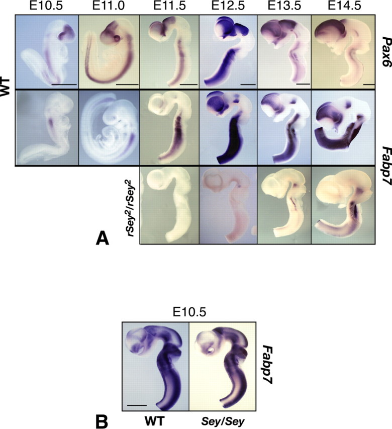
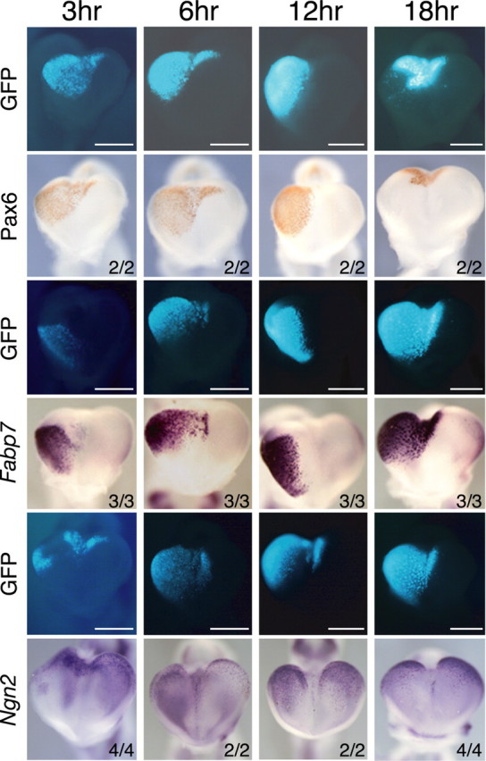
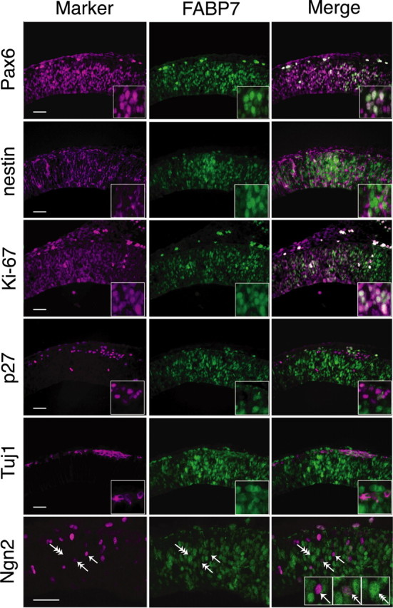
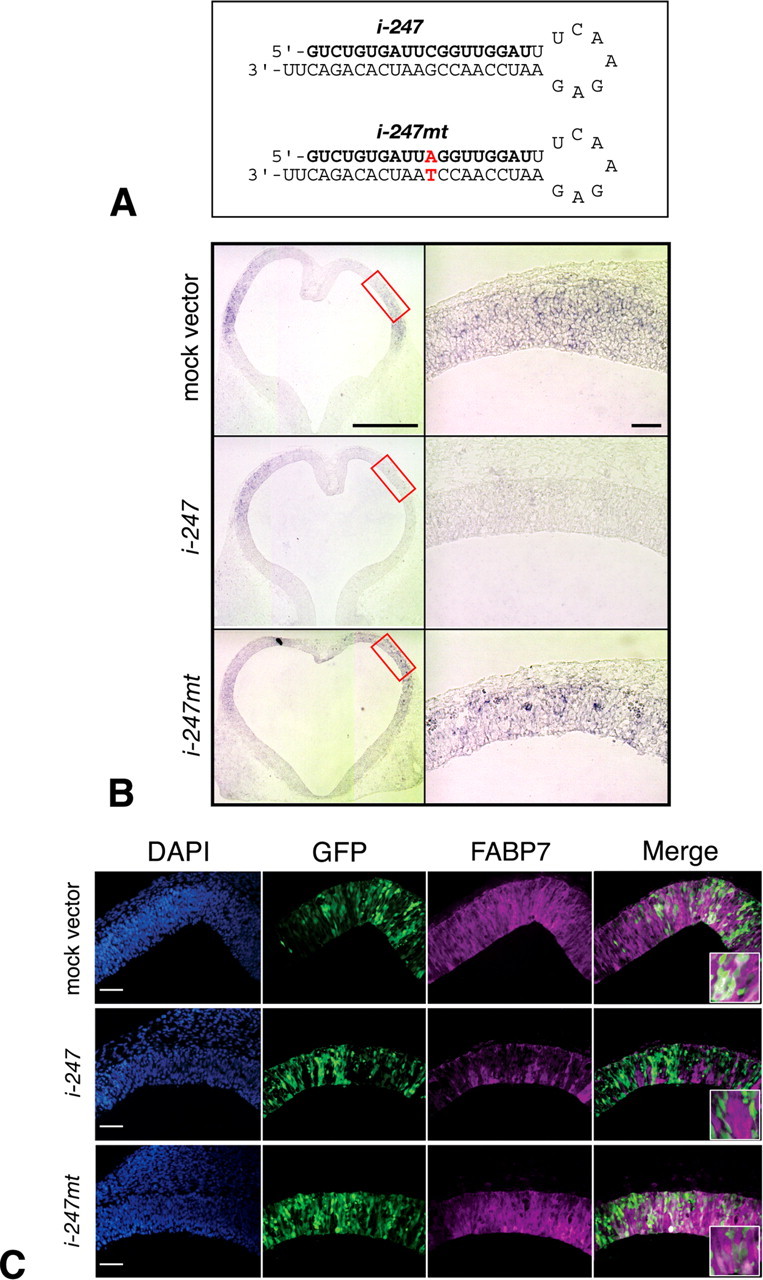
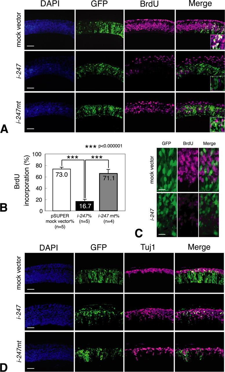
Pax6 is a transcription factor with key functional roles in the developing brain. Pax6 promotes neuronal differentiation via transcriptional regulation of the Neurogenin2 (Ngn2) gene, although Pax6 expression appears in proliferating neuroepithelial cells before the onset of neurogenesis. Here, we identified Fabp7 (BLBP/B-FABP), a member of the fatty acid-binding protein (FABP) family, as a downregulated gene in the embryonic brain of Pax6 mutant rat (rSey2/rSey2) by microarray analysis. Marked reduction of Fabp7 expression was confirmed by quantitative PCR. Spatiotemporal expression patterns of Fabp7 in the wild-type rat embryos from embryonic day 10.5 (E10.5) to E14.5 were similar to those of Pax6, and expression of Fabp7 was undetectable in the rSey2/rSey2 cortex. The expression pattern of Fabp7 in the wild-type mouse embryo at E10.5 (corresponding to E12.5 rat) was different from that in the rat embryo, and no change of expression was observed in the Sey/Sey mouse embryo. Overexpression of exogenous Pax6 mainly induced ectopic expression of Fabp7, rather than of Ngn2, in the early cortical primordium. Interestingly, knocking-down FABP7 function by electroporation of Fabp7 small interfering RNA severely curtailed cell proliferation but promoted neuronal differentiation. We conclude that Fabp7 is a downstream gene of Pax6 transcription factor in the developing rat cortex and essential for maintenance of neuroepithelial cells during early cortical development.


Figures

Figure 1.
Figure 1.
Expression patterns of Pax6 and Fabp7 in the developing rat brain. A, Whole-mount in situ hybridization of the embryo at E10.5 and E11.0 and brain tubes taken from E11.5 to E14.5. At E10.5-E11.0, Pax6 mRNA expression is positive in the forebrain and hindbrain. Fabp7 mRNA expression is positive in the hindbrain. From E12.5 and onwards, expression of Fabp7 becomes evident in the forebrain. Note that the expression pattern of Fabp7 is quite similar to that of Pax6. In rSey2/rSey2 brain, expression of Fabp7 is severely downregulated in the forebrain until E14.5. From E13.5 to E14.5, Fabp7 expression is seen in the rSey2/rSey2 hindbrain. Scale bar, 1 mm. B, Expression patterns of Fabp7 in the developing mouse brain. Whole-mount in situ hybridization of Fabp7 in the brain tube of WT and Sey/Sey at E10.5 (equivalent to rat E12.5). Contrary to the expression in rat embryos, in the mouse, Fabp7 is not specifically expressed in the dorsal telencephalon (Pax6-positive region) but intensely expressed in the ventral telencephalon and in the midbrain (Pax6-negative regions). Also note that Fabp7 expression is almost similar in WT and Sey/Sey brains. Scale bar, 1 mm.
Figure 2.
Figure 2.
Induction of Fabp7 and Ngn2 mRNAs by Pax6 at the early stage of cortical development. pCAX/mPax6 expression vector was coelectroporated with pCAX/GFP into the telencephalic vesicle of E11.5 WT embryos, which were cultured for 3, 6, 12, and 18 h. GFP fluorescence, immunostaining of Pax6 protein, and Fabp7 mRNA are shown in whole mount. At 3 h after electroporation when Pax6 protein was just synthesized, Fabp7 mRNA was immediately induced by Pax6 overexpression. Strong and persistent expression of Fabp7 was observed at least 18 h after electroporation. In contrast, Ngn2 expression was weakly induced at 3 h but not well maintained until 18 h. Numbers represent the number of embryos analyzed. Scale bar, 0.5 mm.
Figure 3.
Figure 3.
FABP7 expression in proliferating NEp cells. Adjacent coronal sections of the WT telencephalon at E12.5. FABP7 expression overlaps with that of Pax6. FABP7-positive cells coexpress progenitor markers, nestin, and Ki-67 but do not express p27 (a nonproliferating marker) or Tuj1 (a neuronal marker). Double immunostaining for FABP7 and Ngn2 showed three types of NEp cells: FABP7 positive/Ngn2 negative (triple arrow), FABP7 moderately positive/Ngn2 moderately positive (double arrow), and FABP7 negative/Ngn2 positive (single arrow). Scale bar, 100 μm.
Figure 4.
Figure 4.
Downregulation of FABP7 expression by siRNA. A, The sequence of the most effective shRNA (i-247) and its mutant (i-247mt) sequences. B, C, pSUPER control mock vector, pSUPER/i-247,or pSUPER/i-247 mt was coelectroporated with pCAX/GFP in the telencephalic vesicle of E11.5 WT rat embryos, which were cultured for 24 h. Reduced expression of Fabp7 mRNA was specifically observed at the region electroporated with pSUPER/i-247 (B, boxes in the left panel), which was not observed in cases electroporated with pSUPER or pSUPER/i-247 mt. Downregulation of FABP7 protein expression was confirmed at the protein level (GFP-positive NEp cells in C). Scale bar, 100 μm.
Figure 5.
Figure 5.
Reduction of FABP7 in neuroepithelial cells is associated with diminished cell proliferation and enhanced neuronal differentiation. A, E11.5 WT rat embryos were electroporated with either control mock vector, pSUPER/i-247,or pSUPER/i-247 mt together with pCAX/GFP in the telencephalic vesicle and cultured for 24 h. Note that immunoreactivity of BrdU is severely reduced in neuroepithelial cells when pSUPER/i-247 was electroporated. B, Quantitative analysis of neuroepithelial cell proliferation. The ordinate shows the percentage of BrdU incorporated cells relative to all GFP-positive cells (ANOVA and Student's t test). C, Morphological changes in pSUPER/i-247 electroporated GFP-positive cells. Cells treated with Fabp7 siRNA become round in shape. Scale bar, 30 μm. D, E11.5 WT rat embryos were electroporated with each expression vector and pCAX/GFP in the telencephalic vesicle and cultured for 48 h. Immunoreactivity of Tuj1 is increased and ectopically observed in the ventricular zone of embryos electroporated with pSUPER/i-247 but not with pSUPER or pSUPER/i-247 mt. Scale bar, 100 μm.

References

    1. Andrews GL, Mastick GS (2003) R-cadherin is a Pax6-regulated, growth-promoting cue for pioneer axons. J Neurosci 23: 9873-9880. - PMC - PubMed
    1. Anthony TE, Klein C, Fishell G, Heintz N (2004) Radial glia serve as neuronal progenitors in all regions of the central nervous system. Neuron 41: 881-890. - PubMed
    1. Anthony TE, Mason HA, Gridley T, Fishell G, Heintz N (2005) Brain lipid-binding protein is a direct target of Notch signaling in radial glial cells. Genes Dev 19: 1028-1033. - PMC - PubMed
    1. Brummelkamp TR, Bernards R, Agami R (2002) A system for stable expression of short interfering RNAs in mammalian cells. Science 296: 550-553. - PubMed
    1. Calegari F, Haubensak W, Haffner C, Huttner WB (2005) Selective lengthening of the cell cycle in the neurogenic subpopulation of neural progenitor cells during mouse brain development. J Neurosci 25: 6533-6538. - PMC - PubMed

Publication types

MeSH terms