

Microsatellite analysis of pleural supernatants could increase sensitivity of pleural fluid cytology
- PMID: 16237222
- PMCID: PMC1888495
- DOI: 10.1016/S1525-1578(10)60583-1
Microsatellite analysis of pleural supernatants could increase sensitivity of pleural fluid cytology
Abstract
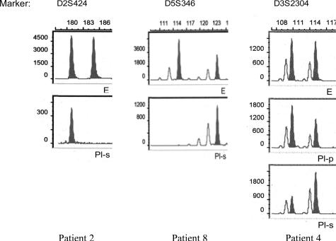
Pleural effusions may result from various inflammatory, hemodynamic, or neoplastic conditions. A common diagnostic problem lies in distinguishing malignant from benign pleural effusions using routine cytological evaluation. We studied pleural fluid samples obtained from 14 patients with histologically confirmed malignancy and from 6 patients with benign pleural effusions using 12 microsatellite markers from 8 different chromosomal regions. Supernatants and cellular sediments of all 20 pleural fluid samples were analyzed. Routine cytological examination was 100% specific for malignancy but was only 57% sensitive. Microsatellite analyses of pleural fluid supernatants showed genetic alterations in tumor patients only. However, 50% of pleural effusions that were considered negative for malignancy by routine cytological analysis showed either loss of heterozygosity or microsatellite instability. The sensitivity of pleural fluid examination rose to 79% when routine cytological assessment was supplemented by molecular studies. Our data suggest that microsatellite analysis increases the sensitivity of cytological pleural fluid examination in assessing potential malignancy and that combining cytological and molecular methods may improve yield and certainty in diagnostically challenging cases.
Figures
Similar articles
-
p53 and FHIT mutations and microsatellite alterations in malignancy-associated pleural effusion.Lung Cancer. 2004 Apr;44(1):33-42. doi: 10.1016/j.lungcan.2003.10.007. Lung Cancer. 2004. PMID: 15013581
-
Can we improve the cytologic examination of malignant pleural effusions using molecular analysis?Ann Thorac Surg. 2005 Oct;80(4):1241-7. doi: 10.1016/j.athoracsur.2005.05.088. Ann Thorac Surg. 2005. PMID: 16181847 Clinical Trial.
-
Microsatellite DNA analysis does not distinguish malignant from benign pleural effusions.Oncol Rep. 2007 Dec;18(6):1507-12. doi: 10.3892/or.18.6.1507. Oncol Rep. 2007. PMID: 17982637
-
Differentiating between malignant and idiopathic pleural effusions: the value of diagnostic procedures.QJM. 2007 Jun;100(6):351-9. doi: 10.1093/qjmed/hcm032. QJM. 2007. PMID: 17525131 Review.
-
Normal volume and cellular contents of pleural fluid.Curr Opin Pulm Med. 2001 Jul;7(4):180-2. doi: 10.1097/00063198-200107000-00002. Curr Opin Pulm Med. 2001. PMID: 11470970 Review.
Cited by
-
Pleural fluid analysis of lung cancer vs benign inflammatory disease patients.Br J Cancer. 2010 Mar 30;102(7):1180-4. doi: 10.1038/sj.bjc.6605607. Epub 2010 Mar 9. Br J Cancer. 2010. PMID: 20216542 Free PMC article.
-
Pleural fluid cell-free DNA integrity index to identify cytologically negative malignant pleural effusions including mesotheliomas.BMC Cancer. 2012 Sep 25;12:428. doi: 10.1186/1471-2407-12-428. BMC Cancer. 2012. PMID: 23009708 Free PMC article.
-
Diagnostic and prognostic value of SHOX2 and SEPT9 DNA methylation and cytology in benign, paramalignant and malignant pleural effusions.PLoS One. 2013 Dec 27;8(12):e84225. doi: 10.1371/journal.pone.0084225. eCollection 2013. PLoS One. 2013. PMID: 24386354 Free PMC article.
-
Molecular testing on serous effusion: An update.Cytojournal. 2021 Dec 6;18:35. doi: 10.25259/Cytojournal_55_2020. eCollection 2021. Cytojournal. 2021. PMID: 35126613 Free PMC article. Review.
-
Cell-free DNA methylation analysis as a marker of malignancy in pleural fluid.Sci Rep. 2024 Feb 5;14(1):2939. doi: 10.1038/s41598-024-53132-x. Sci Rep. 2024. PMID: 38316884 Free PMC article.
References
-
- Light RW. Diagnostic principles in pleural disease. Eur Respir J. 1997;10:476–481. - PubMed
-
- Reithineier A, Lydtin H. Pleural effusion. Internist (Berl) 1996;37:959–968. - PubMed
-
- Garcia-Bonafe M, Moragas A. Differential diagnosis of malignant and reactive cells from serous effusions: image and texture analysis study. Anal Cell Pathol. 1996;12:85–98. - PubMed
-
- Kjellberg SI, Dresler CM, Goldberg M. Pleural cytologies in lung cancer without pleural effusions. Ann Thorac Surg. 1997;64:941–944. - PubMed
Publication types
MeSH terms
Substances
LinkOut - more resources
Full Text Sources
Medical
