. 2006 Jan 20;344(2):315-27.
doi: 10.1016/j.virol.2005.09.010. Epub 2005 Oct 18.

The efficient packaging of Venezuelan equine encephalitis virus-specific RNAs into viral particles is determined by nsP1-3 synthesis

Affiliations

The efficient packaging of Venezuelan equine encephalitis virus-specific RNAs into viral particles is determined by nsP1-3 synthesis

Eugenia Volkova et al. Virology. .

Abstract

Alphaviruses are regarded as attractive systems for expression of heterologous genes and development of recombinant vaccines. Venezuelan equine encephalitis virus (VEE)-based vectors are particularly promising because of their specificity to lymphoid tissues and strong resistance to interferon. To improve understanding of the VEE genome packaging and optimize application of this virus as a vector, we analyzed in more detail the mechanism of packaging of the VEE-specific RNAs. The presence of the RNAs in the VEE particles during serial passaging in tissue culture was found to depend not only on the presence of packaging signal(s), but also on the ability of these RNAs to express in cis nsP1, nsP2 and nsP3 in the form of a P123 precursor. Packaging of VEE genomes into infectious virions was also found to be more efficient compared to that of Sindbis virus, in spite of lower levels of RNA replication and structural protein production.


Figures
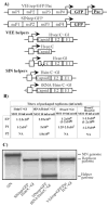
Fig. 1
Fig. 1
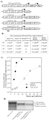
Packaging of VEE and SIN replicons in BHK-21 cells using helpers with deletions of nsP1-4-coding genes. (A) Schematic representation of SIN and VEE replicons and helper genomes. All of the VEE helpers, Hvee/C+Gl, Hvee/C and Hvee/Gl, had natural 5′UTR, derived from VEE TC-83 virus genome, and deletion of nt 520–7290. Hsin/C+Gl and tRNA/Hsin/C+Gl had natural SIN 5′UTR and the 5′ tRNAAsp sequence derived from the naturally occurring SIN DI RNA, respectively. SIN helpers contained a deletion of nt 421–7334 of SIN genome. (B) Titers of packaged VEE and SIN replicons after electroporation and further passaging of the samples. BHK-21 cells were co-transfected with VEErep/GFP/Pac or SINrep/GFP replicons and indicated helper RNAs. Harvested viral particles were used for the next round of infection of naïve BHK-21 cells at the indicated MOIs (measured in inf.u/cell). Viruses were harvested after development of CPE and used for further passaging. Titers refer to unconcentrated virus-containing media. Numbers in the brackets indicate titers in colony-forming units (CFU/ml). All of the experiments were performed multiple times and generated very reproducible titers. NA indicates “not applicable,” because concentration of the packaged replicons after passage 1 was insufficient for infecting cells on the next passage at an MOI of 10 inf.u/cell. (C) Packaging of replicon and helper RNAs into viral particles. 32P-labeled RNAs were isolated from the viral particles released from the cells co-transfected with the indicated replicon and helper RNAs (see Materials and Methods for details). RNA isolated from SIN virus was used as a positive control.
Fig. 2
Fig. 2
Accumulation of the DI RNAs in the samples of VEE TC-83 passaged in BHK-21 cells at high MOI. Passaging was performed as described in Materials and Methods. RNA labeling in the presence of ActD and analysis were performed for the samples harvested after passage 1, 4, 6, 8 and 10 as described in Materials and Methods. Virus-specific RNAs and viral titers are indicated.
Fig. 3
Fig. 3
Packaging of VEE replicons in BHK-21 cells using helpers with deletions of nsP4-coding gene. (A) Schematic representation of VEE replicons and helper genomes. All of the helpers were derived from the VEE TC-83 genome, in which nt 5702–7500 encoding almost the entire nsP4 was deleted. The TGA codon was inserted after nt 5701. Hstop123/C+Gl helper contained an additional insertion of 4 nt sequence after nt 1620. (B) Titers of packaged VEE replicons after electroporation and further passaging of the samples. BHK-21 cells were co-transfected with VEErep/GFP/Pac replicon and indicated helper RNAs. Harvested viral particles were used for the next round of infection of naïve BHK-21 cells at the indicated MOIs (measured in inf.u/cell). Viruses were harvested after the development of CPE and used for further passaging. Titers refer to unconcentrated harvested virus-containing media. NA indicates “not applicable,” because concentration of the packaged replicons after the previous passage was insufficient for infecting cells at an MOI of 10 inf.u/cell. (C) Growth curves of the tri-component genome VEE (VEErep/GFP/Pac + H123/C + H123/Gl) and VEE TC-83. BHK-21 cells were infected at an MOI of 10 inf.u/cell or PFU/cell with tri-component virus and VEE TC-83, respectively. At the indicated times, media were replaced and titers of packaged replicons and virus were determined as described in the Materials and Methods. (D) Packaging of replicon and helper RNAs into viral particles. 32P-labeled RNAs were isolated from the viral particles released from the cells co-transfected with VEErep/GFP/Pac and indicated helper RNAs (see Materials and Methods for details). RNA isolated from VEE TC-83 virus was used as a positive control.
Fig. 4
Fig. 4
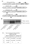
Packaging of VEE replicons in BHK-21 cells using helpers with deletions of nsP4-coding gene. (A) Schematic representation of VEE replicons and helper genomes. All of the helpers were derived from the VEE TC-83 genome, in which nt 5702–7500 were deleted. In all of them, TGA codons were inserted after the last amino acid of nsP3. The TGA codon was also inserted after the last amino acid of nsP1 and nsP2 in H1stop/C+Gl and H12stop/C+Gl, respectively. (B) Packaging of replicon and helper RNAs into viral particles. 32P-labeled RNAs were isolated from the viral particles released from the cells co-transfected with the indicated replicon and helper RNAs (see Materials and Methods for details). (C) Titers of packaged VEE replicons after electroporation and further passaging of the samples. BHK-21 cells were co-transfected with VEErep/GFP/Pac replicon and indicated helper RNAs. Harvested viral particles were used for the next round of infection of naïve BHK-21 cells at the indicated MOIs (measured in inf.u/cell). Viruses were harvested after development of CPE. Titers refer to unconcentrated harvested media. NA indicates “not applicable,” because concentration of the packaged replicons after the previous passage was insufficient for infecting cells at an MOI of 10 inf.u/cell.
Fig. 5
Fig. 5
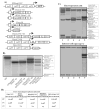
Packaging of SIN replicons in BHK-21 cells using helpers with deletions of the nsP4-coding gene. (A) Schematic representation of SIN replicons and helper genomes. The detailed information is presented in Materials and Methods. (B) Packaging of replicon and helper RNAs into viral particles. 32P-labeled RNAs were isolated from the viral particles released from the cells co-transfected with the indicated replicon and helper RNAs (see Materials and Methods for details). RNA isolated from SIN virus was used as a positive control. (C) Replication of SIN-specific RNAs in BHK-21 cells after electroporation (upper panel) and during passage 1 performed at an MOI 10 inf.u/cell. At 3 h post transfection or post infection, media were replaced by the same media supplemented with dactinomycin (1 μg/ml) and [3H]uridine (20 μCi/ml). After 4 h of incubation at 37°C, RNAs were isolated and analyzed as described in Materials and Methods. SINrep/GFP and Hsin123/C plus Hsin123/Gl helpers, lanes 1; SINrep/GFP and Hsin123/C+Gl helper, lanes 2; SINrep/GFP and Hsin/C plus Hsin/Gl helpers, lanes 3; SINrep/GFP and Hsin/C+Gl helper, lanes 4; SINrep/GFP and tRNA/Hsin/C+Gl helper, lanes 5. (D) Titers of packaged VEE replicons after electroporation and further passaging of the samples. BHK-21 cells were co-transfected with SINrep/GFP replicon and indicated helper RNAs. Harvested viral particles were used for the next round of infection of naïve BHK-21 cells at the indicated MOIs. Viruses were harvested after development of CPE. Titers refer to unconcentrated harvested media. NA indicates “not applicable,” because concentration of the packaged replicons after the previous passage was insufficient for infecting cells at an MOI of 10 inf.u/cell.
Fig. 6
Fig. 6
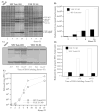
Virus replication and synthesis of virus-specific RNAs in SIN Toto1101- and VEE TC-83-infected cells. (A and B) BHK-21 cells (5 × 10⁵ cells in 35-mm dishes) were infected with SIN Toto1101 or VEE TC-83 at an MOI of 10 PFU/cell. At the indicated times, proteins were pulse-labeled with [35S]methionine as described in Materials and Methods and analyzed on sodium dodecyl sulfate-10% polyacrylamide gels. The gels were dried and autoradiographed (A) or analyzed on a Storm 860 PhoshorImager (B). The levels of synthesis of virus-specific proteins were determined by measuring radioactivity in the protein band corresponding to capsid and were normalized to the number of cysteins and methionines in these proteins. (C and D) BHK-21 cells (5 × 10⁵ cells in 35-mm dishes) were infected with SIN Toto1101 or VEE TC-83 at an MOI of 10 PFU/cell. At the indicated times, media were replaced by the same media supplemented with dactinomycin (1 μg/ml) and [3H]uridine (20 μCi/ml). After 4 h of incubation at 37°C, RNAs were isolated and analyzed as described in Materials and Methods (C). The levels of viral genome replication were determined by excising the bands corresponding to 49S viral genome RNA from the gel shown on panel C, followed by measuring radioactivity by scintillation counting (D). (E) BHK-21 cells (5 × 10⁵ cells in 35-mm dishes) were infected with SIN Toto1101 or VEE TC-83 at an MOI of 10 PFU/cell. At the indicated times, the media were replaced and virus titers were determined as described in Materials and Methods.

Similar articles

Cited by

References

    1. Bredenbeek PJ, Frolov I, Rice CM, Schlesinger S. Sindbis virus expression vectors: Packaging of RNA replicons by using defective helper RNAs. J Virol. 1993;67:6439–6446. - PMC - PubMed
    1. Fayzulin R, Gorchakov R, Petrakova O, Volkova E, Frolov I. Sindbis virus with a tricomponent genome. J Virol. 2005;79(1):637–43. - PMC - PubMed
    1. Frolov I, Schlesinger S. Translation of Sindbis virus mRNA: analysis of sequences downstream of the initiating AUG codon that enhance translation. J Virol. 1996;70(2):1182–90. - PMC - PubMed
    1. Frolova E, Frolov I, Schlesinger S. Packaging signals in alphaviruses. J Virol. 1997;71(1):248–58. - PMC - PubMed
    1. Geigenmuller-Gnirke U, Weiss B, Wright R, Schlesinger S. Complementation between Sindbis viral RNAs produces infectious particles with a bipartite genome. Proc Natl Acad Sci USA. 1991;88:3253–3257. - PMC - PubMed

Publication types

MeSH terms

LinkOut - more resources