. 2005 Oct 21;123(2):347-58.
doi: 10.1016/j.cell.2005.08.004.

The homeodomain transcription factor Irx5 establishes the mouse cardiac ventricular repolarization gradient

Affiliations

The homeodomain transcription factor Irx5 establishes the mouse cardiac ventricular repolarization gradient

Danny L Costantini et al. Cell. .

Abstract

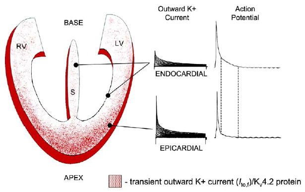
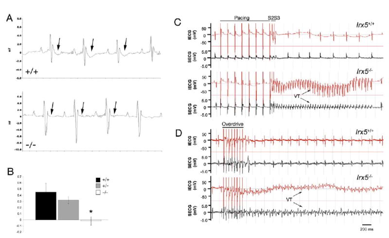
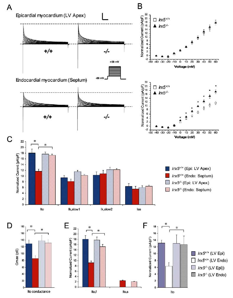
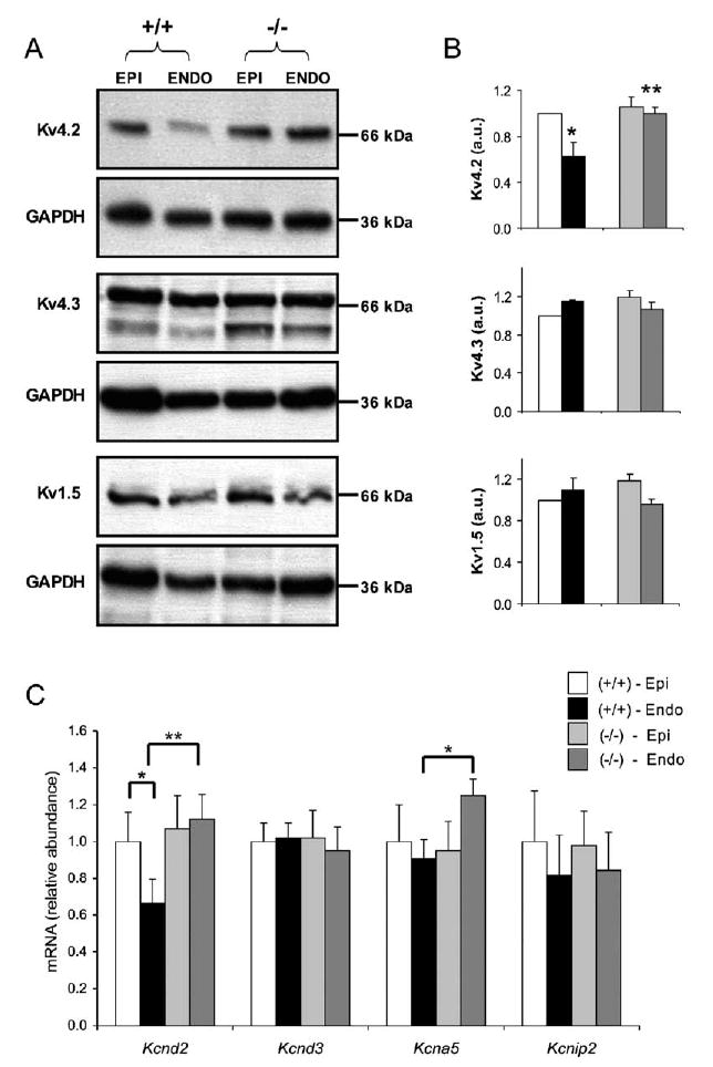
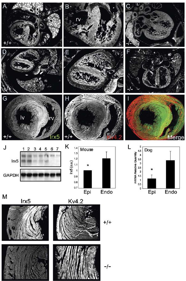
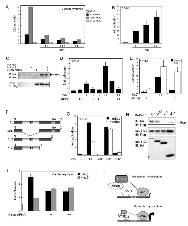
Rhythmic cardiac contractions depend on the organized propagation of depolarizing and repolarizing wavefronts. Repolarization is spatially heterogeneous and depends largely on gradients of potassium currents. Gradient disruption in heart disease may underlie susceptibility to fatal arrhythmias, but it is not known how this gradient is established. We show that, in mice lacking the homeodomain transcription factor Irx5, the cardiac repolarization gradient is abolished due to increased Kv4.2 potassium-channel expression in endocardial myocardium, resulting in a selective increase of the major cardiac repolarization current, I(to,f), and increased susceptibility to arrhythmias. Myocardial Irx5 is expressed in a gradient opposite that of Kv4.2, and Irx5 represses Kv4.2 expression by recruiting mBop, a cardiac transcriptional repressor. Thus, an Irx5 repressor gradient negatively regulates potassium-channel-gene expression in the heart, forming an inverse I(to,f) gradient that ensures coordinated cardiac repolarization while also preventing arrhythmias.


Figures

Figure 1
Figure 1. Repolarization Gradients in the Mammalian Heart
The gradient of density of Ito,f and Kv4.2 protein is shown as red dots on a diagram of the heart. Examples of outward currents and action potential resulting from the high Ito,f/Kv4.2 in epicardial myocardium and low Ito,f/Kv4.2 in endocardial and septal myocardium are shown. See text for details.
Figure 2
Figure 2. Absent T Wave and Inducible Arrhythmias in Irx5−/− Mice
(A) Representative ECGs in the lead II configuration recorded from awake, free-moving mice with the use of telemetric monitoring. Wild-type mice (+/+) show pronounced downward T wave deflections (arrows). No T waves are evident in ECG recordings of Irx5−/− mice (−/−). (B) Quantitation of T wave amplitude (mean ± SEM). n = 6–8; *p < 0.01. (C and D) Representative intracardiac ECG (IECG, red) and surface ECG (SECG, black) in the lead II configuration obtained from wild-type (Irx5+/+) and Irx5−/− mice. (C) Programmed ventricular stimulation at the right ventricular apex using two extra stimuli (“S2S3”) induced episodes of ventricular tachycardia (VT) in Irx5−/− mice, whereas no VTs could be induced in wild-type animals. (D) Rapid overdrive pacing in Irx5−/− mice also induced VTs of long duration.
Figure 3
Figure 3. Shortened Endocardial Action Potentials in Irx5−/− Cardiomyocytes
(A) Representative action-potential traces from Irx5+/+ and Irx5−/−cardiomyocytes from epicardium and endocardium. Irx5−/− endocardial cardiomyocytes demonstrate a shortening of the action potential (arrows). (B) Mean action-potential durations (APD) measured at 25%, 50%, and 90% repolarization following complete depolarization. n = 6–14, *p < 0.05. Scale bars: 20 mV, 25 ms. Data are mean ± SEM.
Figure 4
Figure 4. The Transmural Gradient of Ito Is Eliminated in Irx5−/− Cardiomyocytes
(A) Whole-cell outward K+ currents were recorded from wild-type (+/+) and Irx5−/− (−/−) cardiomyocytes from epicardial (LV apex) and endocardial (septum) regions of the heart. (B) Mean ± SEM normalized peak Ito amplitudes are plotted as a function of test pulse (top, epicardium; bottom, endocardium). (C) Normalized current densities (pA/pF) for Ito, Ik,slow1, Ik,slow2, and Iss measured at +60 mV. (D) Maximum current-conductance values for Ito (Gmax). (E) Normalized current densities (pA/pF) for Ito,f and Ito,s measured at +60 mV. (F) Normalized current densities (pA/pF) for Ito measured in myocytes isolated from LV free wall epicardium or endocardium at +60 mV. For all, n = 6–14, *p < 0.05. Scale bars: 5 nA, 500 ms. Data are mean ± SEM.
Figure 5
Figure 5. Kv4.2 Expression Is Increased in the Endocardium of Irx5−/− Mice
(A) Representative Western blots, using specific anti-Kv4.2, anti-Kv4.3, and anti-Kv1.5 antibodies. (B) Quantitation of Western blot analyses shows increased Kv4.2 protein in Irx5−/− endocardial myocardium. (C) Relative expression of Kcnd2, Kcnd3, Kcna5, and Kcnip2 in the hearts of wild-type and Irx5−/− mice assessed by quantitative real-time RT-PCR. mRNA levels (mean ± SEM) are relative to average wild-type epicardial values; n = 6–8, *p < 0.05 Irx5+/+ endocardial myocardium (Endo) compared with Irx5+/+ epicardial myocardium (Epi), **p < 0.05 Irx5−/− Endo compared with Irx5+/+ Endo. Data are mean ± SEM.
Figure 6
Figure 6. Inverse Gradients of Irx5 and Kv4.2 in the Mouse Heart
(A–F) Immunohistochemistry for Irx5 at E14.5 (A and B) and E16.5 (D and E). Regions in (A) and (D) are magnified in (B) and (E). Only background staining is apparent in Irx5−/−embryos (C and F). lv, left ventricle; rv, right ventricle. (G and H) Irx5 (G) and Kv4.2 (H) expression in adult myocardium. (I) Images in (G) and (H) were pseudocolored green and red, respectively, and digitally merged. (J) Western blot showing Irx5 expression in nuclear extract from epicardial myocardium (lane 1), endocardial myocardium (lane 2), isolated myocytes from epicardial myocardium (lane 3), isolated myocytes from endocardial myocardium (lane 4), isolated neonatal myocytes (lane 5), isolated myocytes from Irx5−/− epicardial myocardium (lane 6), and isolated myocytes from Irx5−/− endocardial myocardium (lane 7). GAPDH is shown as loading control. (K) Quantitation of Irx5 Western blot; n = 3, *p < 0.05. (L) Relative expression of Irx5 mRNA in dog heart; n = 5, *p < 0.05. Data are mean ± SEM. (M) Immunoreactivity of Irx5 and Kv4.2 in the ventricles of adult wild-type (+/+) and Irx5−/−mice (−/−).
Figure 7
Figure 7. Irx5 Directly Represses the Kcnd2 Promoter
(A) Kcnd2 −1094–+592-luciferase and Kcnd2 −432–+592-luciferase (but not Kcnd2 −3162–+592-luciferase) are strongly activated in neonatal cardiac myocytes. Addition of an Irx5 expression construct (Irx5) reduces the activity of Kcnd2 reporters. For this and all other panels: +, 100 ng; ++, 250 ng; +++, 500 ng; ++++, 1000 ng Irx5 expression construct. (B) Irx5 activates Kcnd2-luciferase in COS cells. (C) mBop interacts with Irx4 and Irx5. Immunoprecipitation using anti-HA antibodies followed by immunoblotting against FLAG shows that mBop (arrow) can interact with Irx4 and Irx5. (D) mBop prevents activation of Kcnd2 −1094–+592-luciferase by Irx5. Similar results were obtained with Kcnd2 −432–+592-luciferase. (E) Histone deacetylase inhibition by trichostatin A (TSA) relieves the inhibition of Irx5 activity by mBop. In (D) and (E) for mBop: +, 500 ng; ++, 1000 ng expression constructs. (F) Diagram of Irx5 proteins used in (G) and (H). HD, homeodomain; Iro, Iro box. (G) Irx5ΔHD or Irx5ΔC2 no longer activates transcription, while Irx5ΔC1 activates but is not repressed by mBop. (H) Coimmunoprecipitations show that mBop cannot interact with Irx5ΔC1 or Irx5ΔC2. (I) mBop is required for Irx5-mediated repression in cardiac myocytes. siRNAs against mBop (+, 25 ng; ++, 50 ng) reduced expression of Kcnd2 −432–+592-luciferase and prevented Irx5-mediated repression. Data are mean ± SEM. (J) Model for the role of Irx5; see text for details.

References

    1. Antzelevitch C. Cellular basis and mechanism underlying normal and abnormal myocardial repolarization and arrhythmogenesis. Ann Med. 2004;36 (Suppl 1):5–14. - PubMed
    1. Bao ZZ, Bruneau BG, Seidman JG, Seidman CE, Cepko CL. Irx4 regulates chamber-specific gene expression in the developing heart. Science. 1999;283:1161–1164. - PubMed
    1. Barry DM, Xu H, Schuessler RB, Nerbonne JM. Functional knockout of the transient outward current, long-QT syndrome, and cardiac remodeling in mice expressing a dominant-negative Kv4 alpha subunit. Circ Res. 1998;83:560–567. - PubMed
    1. Bruneau BG. Transcriptional regulation of vertebrate cardiac morphogenesis. Circ Res. 2002;90:509–519. - PubMed
    1. Bruneau BG, Bao ZZ, Tanaka M, Schott JJ, Izumo S, Cepko CL, Seidman JG, Seidman CE. Cardiac expression of the ventricle-specific homeobox gene Irx4 is modulated by Nkx2–5 and dHand. Dev Biol. 2000;217:266–277. - PubMed

Publication types

MeSH terms