. 2006 Jan 15;90(2):619-27.
doi: 10.1529/biophysj.105.061895. Epub 2005 Oct 20.

Photothermal nanotherapeutics and nanodiagnostics for selective killing of bacteria targeted with gold nanoparticles

Affiliations

Photothermal nanotherapeutics and nanodiagnostics for selective killing of bacteria targeted with gold nanoparticles

Vladimir P Zharov et al. Biophys J. .

Abstract

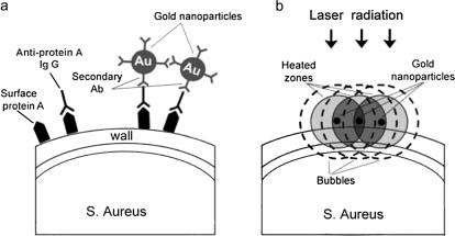
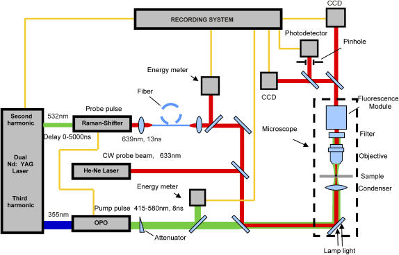
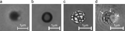
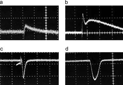
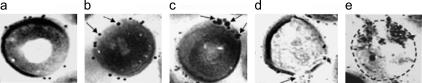
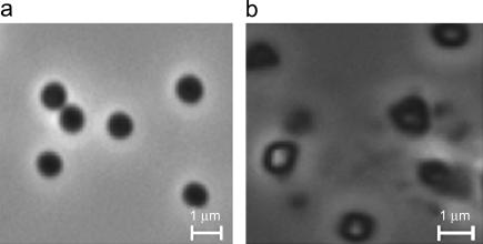
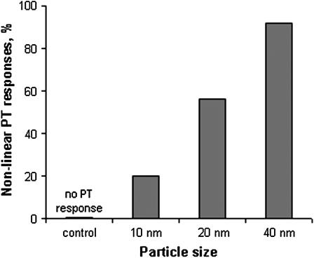
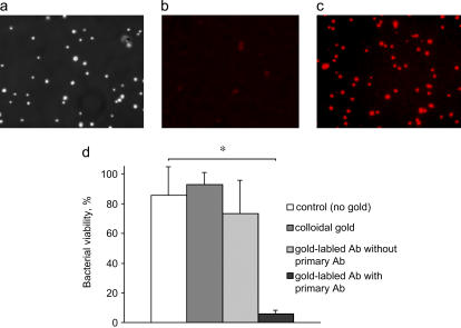
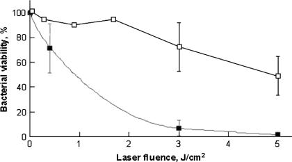
We describe a new method for selective laser killing of bacteria targeted with light-absorbing gold nanoparticles conjugated with specific antibodies. The multifunctional photothermal (PT) microscope/spectrometer provides a real-time assessment of this new therapeutic intervention. In this integrated system, strong laser-induced overheating effects accompanied by the bubble-formation phenomena around clustered gold nanoparticles are the main cause of bacterial damage. PT imaging and time-resolved monitoring of the integrated PT responses assessed these effects. Specifically, we used this technology for selective killing of the Gram-positive bacterium Staphylococcus aureus by targeting the bacterial surface using 10-, 20-, and 40-nm gold particles conjugated with anti-protein A antibodies. Labeled bacteria were irradiated with focused laser pulses (420-570 nm, 12 ns, 0.1-5 J/cm(2), 100 pulses), and laser-induced bacterial damage observed at different laser fluences and nanoparticle sizes was verified by optical transmission, electron microscopy, and conventional viability testing.


Figures

FIGURE 1
FIGURE 1
The principle of selective laser killing of S. aureus targeted with gold nanoparticles.
FIGURE 2
FIGURE 2
Schematics of integrated PT setup for diagnosis and therapy.
FIGURE 3
FIGURE 3
Images of S. aureus with attached gold nanoparticles: (a) phase contrast image (100×, NA 1.3); (b) PT image of bacteria alone (100×, NA 1.25, laser: 520 nm, 100 μJ, with a fluence 20 J/cm2); (c) PT images of bacteria with 40-nm gold particles at laser energy 2 μJ (0.4 J/cm2), time delay between pump and probe laser pulse 30 ns; and (d) PT images of bacteria with 40-nm gold particles at laser energy 10 μJ (2 J/cm2), time delay 120 ns. Dashed lines represent the bacterial boundary in c and d. Arrows in d indicate PT images of single nanoparticles, whereas the arrowhead shows a bubble around one nanocluster.
FIGURE 4
FIGURE 4
Integral PT responses from single S. aureus bacterium at a laser energy of 100 μJ (a) and S. aureus with 40-nm gold nanoparticles at laser energies/fluence of 0.5 μJ/0.1 J/cm2 (b), 3.5 μJ/0.7 J/cm2 (c), and 10 μJ/2 J/cm2 (d). Amplitude (vertical axis)/timescale (horizontal axis) for images ad: 50 mV/400 ns/div; 100 mV/100 ns/div; 200 mV/1 μs/div; and 500 mV/1 μs/div, respectively.
FIGURE 5
FIGURE 5
TEM images of S. aureus conjugated with gold nanoparticles before (a) and after (be) multilaser exposure of 100 pulses, wavelength of 532 nm, and pulse duration of 12 ns at a different conditions (see text): laser fluence of 0.5 J/cm2 and no clusters (b); laser fluence of 0.5 J/cm2 with clustered nanoparticles (c); and laser fluence of 3 J/cm2 at one (d) and several (e) nanocluster numbers. A dashed line represents the bacterial boundary in e. Arrows in b and c indicate penetration of nanoparticles into the wall, and in d, arrows indicate local cell-wall damage.
FIGURE 6
FIGURE 6
Phase-contrast images of S. aureus with 40-nm nanoparticles before (a) and after (b) laser exposure (532 nm; 12 ns; 3 J/cm2, 100 pulses).
FIGURE 7
FIGURE 7
Nonlinear PT response from S. aureus as a function of nanoparticles size. Laser parameters: wavelength, 525 nm; pulse width, 8 ns; and pulse energy, 1 μJ (0.2 J/cm2).
FIGURE 8
FIGURE 8
Selectivity of binding S. aureus with bioconjugated gold nanoparticles. The phase contrast (a) and fluorescent images of S. aureus labeled with chicken anti-goat IgG as secondary antibodies with Alexa Fluor 594 without (b) and with (c) the primary chicken-anti-mouse IgG antibody labeled with Alexa Fluor 488. Bacterial viability was assessed after laser exposure (532 nm, 12 ns, 3 J/cm2) using 40-nm gold nanoparticles by plating the sample on tryptic soy agar (d). The results of plate counting of samples without laser treatment were assumed as 100%. Statistical values were reported as means ± SD of two independent experiments in triplicates. Differences between means were calculated by Student's t-test for paired values. (*p = 0.0006.)
FIGURE 9
FIGURE 9
Efficiency of laser killing of S. aureus targeted with 40-nm gold nanoparticles as a function of laser fluence (0–5 J/cm2). Bacterial viability was assessed by plate counting. Laser parameters are: wavelength, 532 nm; pulse width, 12 ns; and number of pulses, 100. The open squares represent samples irradiated without gold nanoparticles; the solid squares represent samples irradiated with attached gold nanoparticles. The plate-counts of samples without laser treatment were assumed 100% viable. Statistical values are reported as means ± SD of three independent experiments in duplicates.

Similar articles

Cited by

References

    1. Chopra, I. 2003. Antibiotic resistance in Staphylococcus aureus: concerns, causes and cures. Expert Rev. Anti Infect. Ther. 1:45–55. - PubMed
    1. Gemmell, G. C. 2004. Glycopeptide resistance in Staphylococcus aureus: is it a real threat? J. Infect. Chemother. 10:69–75. - PubMed
    1. Novick, R. P. 2003. Autoinduction and signal transduction in the regulation of staphylococcal virulence. Mol. Microbiol. 48:1429–1449. - PubMed
    1. Cheung, A. L., A. S. Bayer, G. Zhang, H. Gresham, and Y. Q. Xiong. 2004. Regulation of virulence determinants in vitro and in vivo in Staphylococcus aureus. FEMS Immunol. Med. Microsc. 40:1–9. - PubMed
    1. Moritz, A., O. Doertbudak, N. Gutknecht, K. Goharkhay, U. Schoop, and W. Sperr. 1997. Nd:YAG laser irradiation of infected root canals in combination with microbiological examinations. J. Am. Dent. Assoc. 128:1525–1530. - PubMed

Publication types