. 2005 Nov 1;102(44):16019-24.
doi: 10.1073/pnas.0508053102. Epub 2005 Oct 20.

Pivotal role of Harakiri in the induction and prevention of gentamicin-induced hearing loss

Affiliations

Pivotal role of Harakiri in the induction and prevention of gentamicin-induced hearing loss

Gilda M Kalinec et al. Proc Natl Acad Sci U S A. .

Abstract

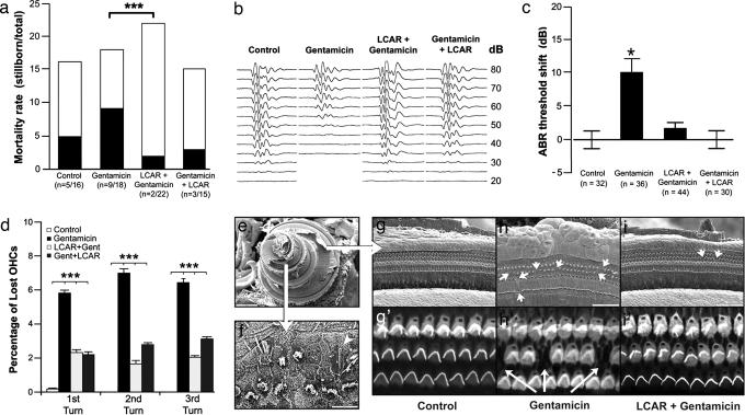
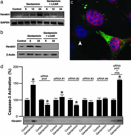
Gentamicin is a widely used ototoxic agent. In this study, we shed light on the mechanisms underlying gentamicin-induced hearing loss. More importantly, we demonstrate in vivo and in vitro the effectiveness of a strategy for preventing drug-induced hearing loss using l-carnitine (LCAR), a safe micronutrient that plays a key role in energy metabolism and detoxification [Rebouche, C. J. & Seim, H. (1998) Annu. Rev. Nutr. 18, 39-61]. We show that LCAR prevents changes in hearing threshold and cochlear damage in newborn guinea pigs exposed to gentamicin in utero. Mechanistically, gentamicin-induced apoptosis of auditory cells is mediated by the extracellular signal-regulated kinase (ERK) 1/2 mitogen-activated protein kinase (MAPK) pathway through up-regulation of the proapoptotic factor Harakiri (Hrk). Most important, small interfering RNA (siRNA) experiments demonstrate that Hrk up-regulation is crucial for gentamicin-induced apoptosis. LCAR, in contrast, prevents both gentamicin-induced Hrk up-regulation and apoptosis acting by means of c-Jun N-terminal kinase (JNK). Together, these results outline pathways for gentamicin-induced hearing loss and its prevention and assign a key role to Hrk in these processes. Thus, our data offer a conceptual framework for designing clinical trials using a safe micronutrient, LCAR, as a simple preventive strategy for iatrogenically induced ototoxicity.


Figures

Fig. 1.
Fig. 1.
Exposure to gentamicin in utero induces, and LCAR prevents, hearing loss and cochlear damage in newborn guinea pigs. (a) LCAR significantly decreases gentamicin-induced neonatal mortality (gentamicin vs. LCAR plus gentamicin, P ≤ 0.003). (b) Typical ABR traces in response to 10-ms clicks. (c) ABR experiments indicate that LCAR prevents the significant shift in hearing threshold associated with exposure to gentamicin. (d) Percentage of lost OHCs in the first, second, and third cochlear turns in newborn guinea pigs. Fourth turn data were not included because of the different nature of the damage (see Results and Discussion and f). (e) SEM image of a full guinea pig cochlea with the smaller, apical fourth turn on the left, and the bigger, basal first turn to the right. (f) Typical appearance of the OHCs in the apical fourth turn of animals exposed to gentamicin. The pattern of distribution is disrupted, many cells are lost, and others show atypical hair bundles. A single giant stereocilia is also visible (arrowhead). (g-i′) SEM and confocal images of more basal turns show the normal structure of the organ of Corti, with three rows of OHCs and scars replacing dead cells (arrows).
Fig. 2.
Fig. 2.
HEI-OC1 cells are sensitive to gentamicin. (a) HEI-OC1 cells quickly incorporate gentamicin, reaching a plateau at 6 h incubation. Gentamicin uptake is not prevented by preincubation with LCAR. (b and c) Gentamicin-filled vesicles (green) accumulate preferentially in the perinuclear region (b), and later distribute in the entire cytoplasm (c). (d and e) Labeling with anti-annexin V antibodies (green) and propidium iodide stain (red) indicates that many gentamicin-exposed cells undergo apoptosis (e), but necrotic (annexin V-negative, propidium iodide-positive) cells were also observed (d). (f and g) SEM studies indicate that plasma membrane blebbing (g) was significantly more frequent in cells exposed to gentamicin than in control cells (f).
Fig. 3.
Fig. 3.
Gentamicin induces, and LCAR prevents, transcriptional up-regulation of Hrk. (a and b) Microarray results were validated by RT-PCR (a) and Western blot (b). (c) Confocal images of HEI-OC1 cells triple-labeled with anti-gentamicin (green), anti-Hrk (red), and the nuclear stain DAPI (blue) show more abundant Hrk expression in cells that incorporate than in those that do not incorporate gentamicin (arrowhead). (d) siRNA experiments confirm that Hrk expression is necessary for gentamicin-induced apoptosis. All of the siRNA oligonucleotides inhibit the expression of Hrk and its up-regulation by gentamicin, and prevent caspase-3 activation. These effects were abolished by cotransfection with Hrk-resistant (rHrk) cDNA.
Fig. 4.
Fig. 4.
Gentamicin-induced up-regulation of Hrk is mediated by MAPKs. (a) Caspase-3 activation experiments indicate that gentamicin-induced apoptosis of HEI-OC1 cells is prevented by preincubation with LCAR and ERK1/2 inhibition, but enhanced by inhibition of JNK. JNK inhibition also interferes with the preventive effect of LCAR. Values are normalized (control = 100%). (b) RT-PCR results confirmed that the effects of the inhibitors of MAPK on caspase-3 activation were associated with changes in Hrk expression. (c) Phosphorylation studies indicate that gentamicin activates ERK1/2 and inactivates JNK. LCAR, in turn, is able to reverse the gentamicin-induced inactivation of JNK. (d-f) Confocal microscopy of HEI-OC1 cells triple-labeled with gentamicin (green), anti-p-ERK (red), and DAPI (blue) confirms that gentamicin activates ERK and demonstrates that gentamicin also induces its translocation to the nucleus. A cell with numerous gentamicin-filled vesicles in the cytoplasm and p-ERK concentrated in the nucleus is pointed out with an arrow. An arrowhead indicates a cell that incorporates only a small amount of gentamicin and shows cytoplasmic labeling with anti-p-ERK.

Similar articles

Cited by

References

    1. Chambers, H. F. & Sande, M. A. (1996) in Goodman & Gilman's The Pharmacological Basis of Therapeutics, eds. Hardman, J. G., Limbird, L. E., Molinoff, P. B., Ruddon, R. W. & Gilman, A. G. (McGraw-Hill, New York), pp. 1103-1121.
    1. Romero, R., Espinoza, J., Chaiworapongsa, T. & Kalache, K. (2002) Semin. Neonatol. 7, 259-274. - PubMed
    1. Henley, C. M. & Rybak, L. P. (1995) Brain Res. Brain Res. Rev. 20, 68-90. - PubMed
    1. Priuska, E. M. & Schacht, J. (1995) Biochem. Pharmacol. 50, 1749-1752. - PubMed
    1. Forge, A. & Schacht, J. (2000) Audiol. Neurootol. 5, 3-22. - PubMed

Publication types