. 2005 Oct 25;102(43):15599-604.
doi: 10.1073/pnas.0409730102. Epub 2005 Oct 20.

Creating oral squamous cancer cells: a cellular model of oral-esophageal carcinogenesis

Affiliations

Creating oral squamous cancer cells: a cellular model of oral-esophageal carcinogenesis

Gitta Goessel et al. Proc Natl Acad Sci U S A. .

Abstract

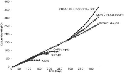
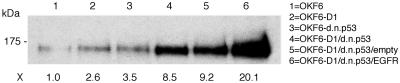
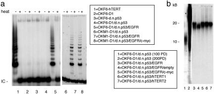
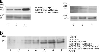
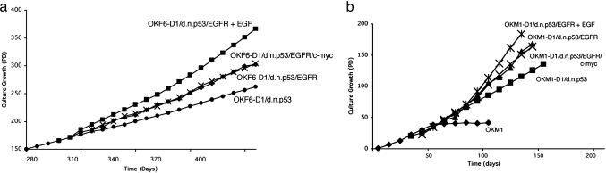
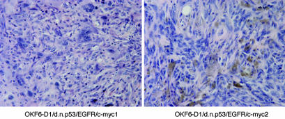
Immortalization and malignant transformation are important steps in tumor development. The ability to induce these processes from normal human epithelial cells with genetic alterations frequently found in the corresponding human cancer would significantly enhance our understanding of tumor development. Alterations in several key intracellular regulatory pathways (the pRB, p53, and mitogenic signaling pathways and the telomere maintenance system) appear to be sufficient for the neoplastic transformation of normal human cells. Nevertheless, in vitro transformation models to date depend on viral oncogenes, most prominently the simian virus 40 early region, to induce immortalization and malignant transformation of normal human epithelial cells. Here, we demonstrate a transformation model creating oral-esophageal cancer cells by using a limited set of genetic alterations frequently observed in the corresponding human cancer. In a stepwise model, cyclin D1 overexpression and p53 inactivation led to immortalization of oral keratinocytes. Additional ectopic epithelial growth factor receptor overexpression followed by c-myc overexpression as well as consecutive reactivation of telomerase induced by epithelial growth factor receptor sufficed to transform oral epithelial cells, truly recapitulating the development of the corresponding human disease.


Figures

Fig. 1.
Fig. 1.
Replicative life span of parental and derived oral keratinocytes. Growth characteristics of OKF6, OKF6-D1, OKF6-dnp53, OKF6-D1/dnp53, OKF6-D1/dnp53/EGFR, and OKF6-D1/dnp53/EGFR plus EGF. The replicative life span was assessed by calculating the PDs of each cell line. Immortalization was assessed if cells grew at least three times beyond the life span of the parental cells.
Fig. 2.
Fig. 2.
Effects of EGFR overexpression in the generated oral keratinocytes. Equal amounts of protein for OKF6, OKF6-D1, OKF6-dnp53, OKF6-D1/dnp53, OKF6-D1/dnp53/empty, and OKF6-D1/dnp53/EGFR were separated on a 6% SDS/PAGE. The level of EGFR overexpression was compared by subsequent immunoblot analysis using a polyclonal EGFR antibody (1005). EGFR overexpression is indicated as fold increase compared to parental OKF6 cells.
Fig. 3.
Fig. 3.
Telomerase activity and telomere length in parental and derived oral keratinocytes. (a) Cellular extracts (100 ng) of OKF6-hTERT, OKF6-D1, OKF6-dnp53, OKF6-D1/dnp53, OKF6-D1/dnp53/EGFR, as well as OKM1-D1/dnp53, OKM1-D1/dnp53/EGFR, and OKM1-D1/dnp53/EGFR/c-myc cells were assayed for telomerase activity using the PCR-based TRAP assay. Heat-treated (heat) samples served as negative control, and OKF6-hTERT served as a positive control. IC is an internal PCR control. (b) Telomere length for preimmortal OKF6-D1/dnp53 (100 PD), immortal OKF6-D1/dnp53 (200 PD), OKF6-D1/dnp53/EGFR, OKF6-D1/dnp53/EGFR/empty, and OKF6-D1/dnp53/EGFR/c-myc cells was analyzed by hybridization of genomic DNA with a specific oligonucleotide probe. Two different clones of OKF6-D1/dnp53/hTERT cells served as positive control.
Fig. 4.
Fig. 4.
Western Blot analysis of AKT/phospho-AKT, ERK/phospho-ERK as well as c-myc in the generated oral keratinocytes. (a) Equal amounts of protein of OKF6-D1/dnp53, OKF6-D1/dnp53/empty, and OKF6-D1/dnp53/EGFR cells were separated with 10% SDS/PAGE, and a Western Blot was performed. The primary antibodies used were polyclonal AKT1/PKBa, monoclonal phospho-AKT (Ser-473), polyclonal ERK1/2 (C-14), and monoclonal phospho-ERK1/2 (E-4). (b) Equal amounts of protein for OKF6, OKF6-D1, OKF6-D1/dnp53, OKF6-D1/dnp53/EGFR, OKF6-D1/dnp53/EGFR/c-myc (two individual clones), and TE-12 were separated with 10% SDS/PAGE. The level of c-myc overexpression was compared by immunoblot analysis using a polyclonal c-myc antibody (N-262).
Fig. 5.
Fig. 5.
Replicative life span of oral keratinocytes additionally overexpressing EGFR and c-myc. Growth characteristics of OKF6-D1/dnp53, OKF6-D1/dnp53/EGFR, OKF6-D1/dnp53/EGFR plus EGF, and OKF6-D1/dnp53/EGFR/c-myc cells (a), as well as OKM1, OKM1-D1/dnp53, OKM1-D1/dnp53/EGFR, OKM1-D1/dnp53/EGFR plus EGF, and OKM1-D1/dnp53/EGFR/c-myc cells (b) are displayed. The replicative life span was assessed by calculating the PD values.
Fig. 6.
Fig. 6.
Nude mouse tumors induced by generated oral cancer cells. Tumors induced by the generated human oral cancer cells contain histologic features of poorly differentiated squamous cell cancer mixed with spindle cells. Depicted are representative photomicrographs of tumors from OKF6-D1/dnp53/EGFR/c-myc (two individual clones) shown with hematoxylin/eosin staining. (Magnification, ×400.)

References

    1. Hayflick, L. (1965) Exp. Cell Res. 37, 614–636. - PubMed
    1. Harada, H., Nakagawa, H., Oyama, K., Takaoka, M., Andl, C. D., Jacobmeier, B., von Werder, A., Enders, G. H., Opitz, O. G. & Rustgi, A. K. (2003) Mol. Cancer Res. 1, 729–738. - PubMed
    1. Reddel, R. R. (2000) Carcinogenesis 21, 477–484. - PubMed
    1. Hahn, W. C. & Weinberg, R. A. (2002) Nat. Rev. Cancer 2, 331–341. - PubMed
    1. Bartkova, J., Lukas, J., Muller, H., Strauss, M., Gusterson, B. & Bartek, J. (1995) Cancer Res. 55, 949–956. - PubMed

Publication types

MeSH terms