

Complement-induced regulatory T cells suppress T-cell responses but allow for dendritic-cell maturation
- PMID: 16239430
- PMCID: PMC1895395
- DOI: 10.1182/blood-2005-07-2951
Complement-induced regulatory T cells suppress T-cell responses but allow for dendritic-cell maturation
Abstract
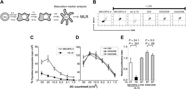
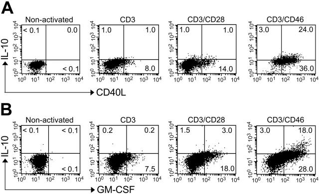
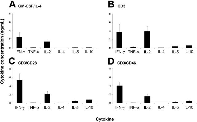
Concurrent activation of the T-cell receptor (TCR) and complement regulator CD46 on human CD4+ T lymphocytes induces Tr1-like regulatory T cells that suppress through IL-10 secretion bystander T-cell proliferation. Here we show that, despite their IL-10 production, CD46-induced T-regulatory T cells (Tregs) do not suppress the activation/maturation of dendritic cells (DCs). DC maturation by complement/CD46-induced Tregs is mediated through simultaneous secretion of GM-CSF and soluble CD40L, factors favoring DC differentiation and reversing inhibitory effects of IL-10. Thus, CD46-induced Tregs produce a distinct cytokine profile that inhibits T-cell responses but leaves DC activation unimpaired. Such "DC-sparing" Tregs could be desirable at host/environment interfaces such as the gastrointestinal tract where their specific cytokine profile provides a mechanism that ensures unresponsiveness to commensal bacteria while maintaining reactivity to invading pathogens.
Figures


Similar articles
-
IL-1 beta enhances CD40 ligand-mediated cytokine secretion by human dendritic cells (DC): a mechanism for T cell-independent DC activation.J Immunol. 2002 Jan 15;168(2):713-22. doi: 10.4049/jimmunol.168.2.713. J Immunol. 2002. PMID: 11777965 Clinical Trial.
-
Tumor necrosis factor alpha and CD40 ligand antagonize the inhibitory effects of interleukin 10 on T-cell stimulatory capacity of dendritic cells.Cancer Res. 2000 Aug 15;60(16):4485-92. Cancer Res. 2000. PMID: 10969796
-
CD46 engagement on human CD4+ T cells produces T regulatory type 1-like regulation of antimycobacterial T cell responses.Infect Immun. 2010 Dec;78(12):5295-306. doi: 10.1128/IAI.00513-10. Epub 2010 Oct 4. Infect Immun. 2010. PMID: 20921150 Free PMC article.
-
Induction of Interleukin-10 Producing Dendritic Cells As a Tool to Suppress Allergen-Specific T Helper 2 Responses.Front Immunol. 2018 Mar 19;9:455. doi: 10.3389/fimmu.2018.00455. eCollection 2018. Front Immunol. 2018. PMID: 29616018 Free PMC article. Review.
-
CD40 and dendritic cell function.Crit Rev Immunol. 2003;23(1-2):83-107. doi: 10.1615/critrevimmunol.v23.i12.50. Crit Rev Immunol. 2003. PMID: 12906261 Review.
Cited by
-
CD4+ Foxp3+ T cells promote aberrant immunoglobulin G production and maintain CD8+ T-cell suppression during chronic liver disease.Hepatology. 2017 Feb;65(2):661-677. doi: 10.1002/hep.28894. Epub 2016 Dec 19. Hepatology. 2017. PMID: 27774611 Free PMC article.
-
Expansion and characteristics of human T regulatory type 1 cells in co-cultures simulating tumor microenvironment.Cancer Immunol Immunother. 2007 Sep;56(9):1429-42. doi: 10.1007/s00262-007-0280-9. Epub 2007 Jan 31. Cancer Immunol Immunother. 2007. PMID: 17265021 Free PMC article.
-
Assessment of seasonal influenza A virus-specific CD4 T-cell responses to 2009 pandemic H1N1 swine-origin influenza A virus.J Virol. 2010 Apr;84(7):3312-9. doi: 10.1128/JVI.02226-09. Epub 2010 Jan 13. J Virol. 2010. PMID: 20071564 Free PMC article.
-
Toll-like receptors and immune regulation: their direct and indirect modulation on regulatory CD4+ CD25+ T cells.Immunology. 2007 Oct;122(2):149-56. doi: 10.1111/j.1365-2567.2007.02651.x. Immunology. 2007. PMID: 17848162 Free PMC article. Review.
-
Targeting the Complement Alternative Pathway Permits Graft Versus Leukemia Activity while Preventing Graft Versus Host Disease.Clin Cancer Res. 2020 Jul 1;26(13):3481-3490. doi: 10.1158/1078-0432.CCR-19-1717. Epub 2020 Jan 9. Clin Cancer Res. 2020. PMID: 31919135 Free PMC article.
References
-
- Sakaguchi S. Regulatory T-cells: key controllers of immunologic self-tolerance. Cell. 2000;101: 455-458. - PubMed
-
- Shevach EM. Regulatory T-cells in autoimmunity. Annu Rev Immunol. 2000;18: 423-449. - PubMed
-
- Bluestone JA, Abbas AK. Natural versus adaptive regulatory T-cells. Nat Rev Immunol. 2003;3: 253-257. - PubMed
-
- Jonuleit H, Schmitt E. The regulatory T-cell family: distinct subsets and their interrelations. J Immunol. 2003;171: 6323-6327. - PubMed
-
- Fehérvari Z, Sakaguchi S. Development of CD25+CD4+ regulatory T-cells. Curr Opin Immunol. 2004;16: 203-208. - PubMed
Publication types
MeSH terms
Substances
Grants and funding
LinkOut - more resources
Full Text Sources
Other Literature Sources
Research Materials
