. 2005 Nov;73(11):7180-9.
doi: 10.1128/IAI.73.11.7180-7189.2005.

Merozoite surface antigen 2 proteins of Babesia bovis vaccine breakthrough isolates contain a unique hypervariable region composed of degenerate repeats

Affiliations

Merozoite surface antigen 2 proteins of Babesia bovis vaccine breakthrough isolates contain a unique hypervariable region composed of degenerate repeats

Shawn J Berens et al. Infect Immun. 2005 Nov.

Abstract

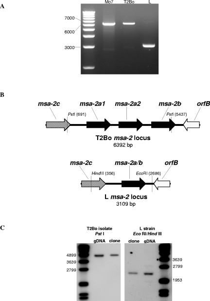
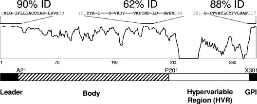
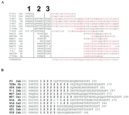
The merozoite surface antigen 2 (MSA-2) proteins of Babesia bovis are members of the variable merozoite surface antigen (VMSA) family that have been implicated in erythrocyte invasion and are important targets for antibody-mediated blocking of invasion. Extensive sequence variation in another VMSA member, MSA-1, has been shown in all vaccine breakthrough isolates. To test the hypothesis that the msa-2 genes of vaccine breakthrough isolates would also encode a diverse set of proteins, the complete msa-2 locus was characterized from 12 Australian B. bovis strains and isolates, including two vaccine strains and eight vaccine breakthrough isolates, and compared to the loci in previously and newly characterized American strains. In contrast to American strains, the msa-2 loci of all Australian strains and isolates examined contain, in addition to msa-2c, only a solitary gene (designated msa-2a/b) closely related to American strain msa-2a and msa-2b. Nevertheless, the proteins encoded by these genes are quite diverse both between and within geographic regions and harbor evidence of genetic exchange among other VMSA family members, including msa-1. Moreover, all but one of the Australian breakthrough isolate MSA-2a/b proteins is markedly different from the vaccine strain from which immune escape occurred, consistent with their role in strain-specific protective immunity. The densest distribution of polymorphisms occurs in a hypervariable region (HVR) within the carboxy third of the molecule that is highly proline rich. Variation in length and content of the HVR is primarily attributable to differences in the order and number of degenerate nucleotide repeats encoding three motifs of unknown function.


Figures

FIG. 1.
FIG. 1.
The Australian virulent B. bovis L strain contains a single msa-2a/b gene. (A) Ethidium bromide-stained agarose gel of the msa-2 locus amplified from biological clone Mo7 (lane 2), Texas T2Bo strain (lane 3), and Australian virulent L strain (lane 4) with a 1.0-kb marker in lane 1. (B) Schematic of the msa-2 locus from T2Bo, based on the sequence of BAC clone 1K11 (see Materials and Methods), and L, based on the nucleotide sequence of the amplified msa-2 locus. (C) Southern blot of T2Bo genomic DNA and a T2Bo msa-2 locus clone digested with PstI and probed with digoxigenin-labeled T2Bo msa-2a/b-specific oligonucleotide (left panel) and L strain genomic DNA and L msa-2 locus clone digested with EcoRI/HindIII and probed with digoxigenin-labeled L msa-2a/b-specific oligonucleotide (right panel). Probe detection was by chemiluminescence. Size markers are in the far left and far right lanes, and the relevant markers are designated in base pairs to the left and right of the panels.
FIG. 2.
FIG. 2.
The MSA-2a/b proteins are diverse and have a hypervariable region in the carboxy half of the molecule. An alignment similarity plot of all examined MSA-2a/b proteins is shown. Regions of relative high identity are indicated by an overlying bar with sequence information and composite percent identity provided. Dashes in sequences indicate nonconserved residues. A schematic of MSA-2a/b molecular organization based on sequence variation and known function is shown below. Protein residues noted at the junction of sequence segments are initiating residues.
FIG. 3.
FIG. 3.
Vaccine breakthrough isolate MSA-2a/b protein sequences differ from their associated vaccine strains. (A) MSA-2a/b sequence alignment of the Australian T vaccine strain compared to the vaccine breakthrough G isolates (G52, G51, G36, and G06). (B) MSA-2a/b sequence alignment of the Australian K vaccine strain compared to the vaccine breakthrough F isolates (F3, F28, F35, and F40). The underlined sequences indicate stretches of complete conservation. (C) MSA-2a/b amino-terminal region alignment of T vaccine strain T-1, vaccine breakthrough isolate G06, and Mo7 2a1. Boxes highlight regions of high identity between G06 and Mo7 2a1. Spaces denote identity, and dashes indicate gaps. Alignments were constructed by CLUSTALW.
FIG. 4.
FIG. 4.
The HVR of MSA-2a/b contains a series of three, proline-rich, repeating motifs. (A) Amino acid alignment of all 22 MSA-2a/b proteins examined. A period replaces all nonproline residues, a red “X” replaces all proline residues, and dashes indicate gaps in sequences. (B) Manual amino acid alignment of 10 representative MSA-2a/b HVRs along with the 3′ region upstream of the GPI anchor signal sequence of F35, F40, and R1A MSA-1 and F40 and F28 MSA-2c. Alignments are based on both amino acid and nucleotide sequences. Motifs are highlighted by color. Sequences of the first motif are red, sequences of the second motif are purple, sequences of the third motif are green, and the common GNLNG sequence is blue. Residues that are not assigned to a motif are in black letters. Blank spaces indicate gaps. A black bisected underline splits the HVR into amino and carboxy regions as labeled.
FIG. 5.
FIG. 5.
The amino-terminal region of the HVR is encoded by a series of degenerate nucleotide repeats. (A) Nucleotide sequence alignment of representative segments from the amino-terminal region (Fig. 4B) of HVRs. Base pair substitutions in reference to the consensus sequence are italicized; base pairs identical with the consensus sequence are red. The encoded amino acid sequences of these segments, with their relative positions in the 5′ region of the HVR, are aligned in the left half of the figure. The amino acid alignment is partitioned into three segments and numbered as indicated at the top of the alignment. “Md” indicates sequences derived from the middle portion of the amino half of the HVR region. Spaces denote gaps. (B) Sequences of the of representative HVRs with transformation of the amino acid sequence fragments of the 5′ region with the segment number to which their nucleotide sequences mapped.
FIG. 6.
FIG. 6.
The HVR has sequence similarities to other VMSA family members. Three separate amino acid alignments are shown. Sequences shared among VMSA family members are boldfaced. The bracketed segments highlight motifs 1 and 3 as specified in the text (Fig. 4). Dashes indicate sequence gaps. The fractions listed at the right of the sequences are the frequency with which the representative sequence segments are present in the indicated VMSA family members that were examined.

References

    1. Acosta-Serrano, A., E. Vassella, M. Liniger, C. Kunz Renggli, R. Brun, I. Roditi, and P. T. Englund. 2001. The surface coat of procyclic Trypanosoma brucei: programmed expression and proteolytic cleavage of procyclin in the tsetse fly. Proc. Natl. Acad. Sci. USA 98:1513-1518. - PMC - PubMed
    1. Anziani, O. S., A. A. Guglielmone, A. A. Abdala, D. H. Aguirre, and A. J. Mangold. 1993. Acquired protection in Frisian steers with Babesia bovis attenuated vaccine. Rev. Med. Vet. 74:47-49.
    1. Ayala, F. J., A. A. Escalante, and S. M. Rich. 1999. Evolution of Plasmodium and the recent origin of the world populations of Plasmodium falciparum. Parasitologia 41:55-68. - PubMed
    1. Bock, R. E., A. J. de Vos, T. G. Kingston, I. A. Shiels, and R. J. Dalgliesh. 1992. Investigations of breakdowns in protection provided by living Babesia bovis vaccine. Vet. Parasitol. 43:45-56. - PubMed
    1. Bock, R. E., A. J. de Vos, A. Lew, T. G. Kingston, and I. R. Fraser. 1995. Studies on failure of T strain live Babesia bovis vaccine. Aust. Vet. J. 72:296-300. - PubMed

Publication types

MeSH terms

Associated data

LinkOut - more resources