. 2005 Nov;73(11):7236-42.
doi: 10.1128/IAI.73.11.7236-7242.2005.

Identification of gmhA, a Yersinia pestis gene required for flea blockage, by using a Caenorhabditis elegans biofilm system

Affiliations

Identification of gmhA, a Yersinia pestis gene required for flea blockage, by using a Caenorhabditis elegans biofilm system

Creg Darby et al. Infect Immun. 2005 Nov.

Abstract

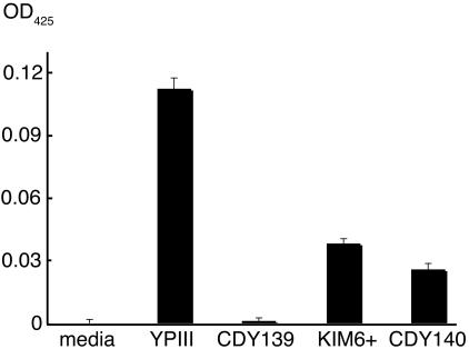
Yersinia pestis, the cause of bubonic plague, blocks feeding by its vector, the flea. Recent evidence indicates that blockage is mediated by an in vivo biofilm. Y. pestis and the closely related Yersinia pseudotuberculosis also make biofilms on the cuticle of the nematode Caenorhabditis elegans, which block this laboratory animal's feeding. Random screening of Y. pseudotuberculosis transposon insertion mutants with a C. elegans biofilm assay identified gmhA as a gene required for normal biofilms. gmhA encodes phosphoheptose isomerase, an enzyme required for synthesis of heptose, a conserved component of lipopolysaccharide and lipooligosaccharide. A Y. pestis gmhA mutant was constructed and was severely defective for C. elegans biofilm formation and for flea blockage but only moderately defective in an in vitro biofilm assay. These results validate use of the C. elegans biofilm system to identify genes and pathways involved in Y. pestis flea blockage.


Figures

FIG. 1.
FIG. 1.
Nematode growth on Y. pestis and Y. pseudotuberculosis strains. The percentage of L4 C. elegans was determined 2 days after eggs were laid on lawns of the indicated strains. gmhA genotypes: +, wild type; −, chromosomal deletion; −/+, deletion strain carrying complementing plasmid pCBD32; −/v, deletion strain carrying vector. Data are means and standard deviations from three independent experiments for each strain except YPIII, which was assayed six times.
FIG. 2.
FIG. 2.
Biofilms on C. elegans made by Yersinia wild-type and gmhA mutant strains. A nematode grown on E. coli food strain OP50 is shown for comparison. Black arrows, mouth. Wild-type Y. pestis and Y. pseudotuberculosis biofilms cover the mouth completely and block feeding. Most Y. pestis gmhA biofilms fail to attach to the mouth (not shown), but a few are as shown, accounting for the 2% of C. elegans animals that fail to grow normally on this strain (Fig. 1). Y. pseudotuberculosis gmhA biofilms attach to the side of the head posterior to the mouth; white arrows mark the forward edge of the biofilm.
FIG. 3.
FIG. 3.
LOS and LPS of Yersinia mutants. (A) Core oligosaccharide of gmhA mutants migrates faster through gels, indicating truncation. +, wild type; −, gmhA deletion mutant; −/+, mutant complemented with pCBD32. (B) O antigen is absent in the Y. pseudotuberculosis gmhA mutant and in strains with mutations in known O-antigen genes. Wild-type YPIII (+) shows a typical ladder-like pattern produced by variable numbers of O-antigen repeat units. Faint bands in mutant samples are presumed protease-resistant proteins.
FIG. 4.
FIG. 4.
The Y. pestis gmhA mutant is defective for flea blockage. (A) Colonization of flea digestive tract after a single blood meal containing Y. pestis. (B) Flea blockage during the 4-week period after an infected blood meal. The 25% level of blockage for the wild type is typical for strain KIM6+. (C) Y. pestis CFU taken up in the infectious blood meal (day 0) and in infected fleas 7 and 28 days postinfection. Data are means and standard deviations from two independent experiments. w.t., wild type.
FIG. 5.
FIG. 5.
In vitro biofilms. Bacteria were grown standing for 4 days in polystyrene dishes, after which broth and planktonic cells were removed and adherent biofilm matrix was detected with WGA conjugated to alkaline phosphatase (see Materials and Methods for details). Data are means and standard errors of the means for four replicates in a single experiment; results are typical of multiple experiments. OD425, optical density at 425 nm.

References

    1. Achtman, M., K. Zurth, G. Morelli, G. Torrea, A. Guiyoule, and E. Carniel. 1999. Yersinia pestis, the cause of plague, is a recently emerged clone of Yersinia pseudotuberculosis. Proc. Natl. Acad. Sci. USA 96:14043-14048. - PMC - PubMed
    1. Bateman, A., E. Birney, L. Cerruti, R. Durbin, L. Etwiller, S. R. Eddy, S. Griffiths-Jones, K. L. Howe, M. Marshall, and E. L. Sonnhammer. 2002. The Pfam protein families database. Nucleic Acids Res. 30:276-280. - PMC - PubMed
    1. Brooke, J. S., and M. A. Valvano. 1996. Biosynthesis of inner core lipopolysaccharide in enteric bacteria: identification and characterization of a conserved phosphoheptose isomerase. J. Biol. Chem. 271:3608-3614. - PubMed
    1. Darby, C., J. W. Hsu, N. Ghori, and S. Falkow. 2002. Caenorhabditis elegans: plague bacteria biofilm blocks food intake. Nature 417:243-244. - PubMed
    1. Deng, W., V. Burland, G. Plunkett III, A. Boutin, G. F. Mayhew, P. Liss, N. T. Perna, D. J. Rose, B. Mau, S. Zhou, D. C. Schwartz, J. D. Fetherston, L. E. Lindler, R. R. Brubaker, G. V. Plano, S. C. Straley, K. A. McDonough, M. L. Nilles, J. S. Matson, F. R. Blattner, and R. D. Perry. 2002. Genome sequence of Yersinia pestis KIM. J. Bacteriol. 184:4601-4611. - PMC - PubMed

Publication types

MeSH terms

LinkOut - more resources