. 2005 Nov;73(11):7243-51.
doi: 10.1128/IAI.73.11.7243-7251.2005.

Studies of fibronectin-binding proteins of Streptococcus equi

Affiliations

Studies of fibronectin-binding proteins of Streptococcus equi

Jonas Lannergård et al. Infect Immun. 2005 Nov.

Abstract

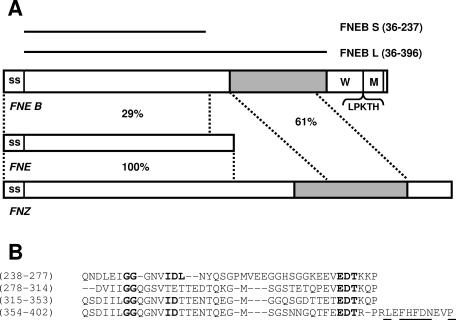
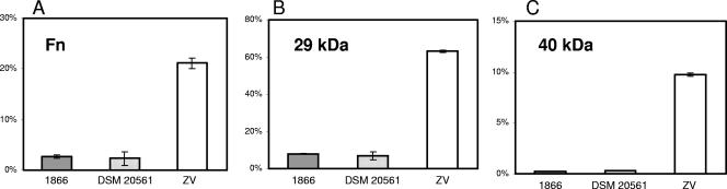
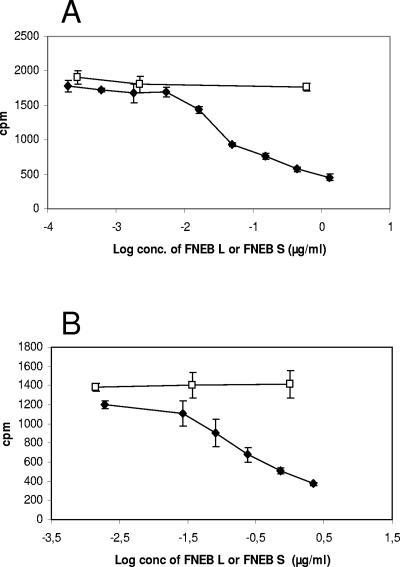
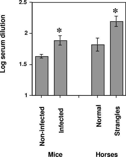
Streptococcus equi subsp. equi is the causative agent of strangles, a disease of the upper respiratory tract in horses. The initiation of S. equi subsp. equi infection is likely to involve cell surface-anchored molecules mediating bacterial adhesion to the epithelium of the host. The present study describes the cloning and characterization of FNEB, a fibronectin-binding protein with cell wall-anchoring motifs. FNEB can thus be predicted as cell surface located, contrary to the two previously characterized fibronectin-binding proteins in S. equi subsp. equi, FNE and SFS. Assays of antibody titers in horses and in experimentally infected mice indicate that the protein is immunogenic and expressed in vivo during S. equi subsp. equi infection. Using Western ligand blotting, it was shown that FNEB binds to the N-terminal 29-kDa fragment of fibronectin, while SFS and FNE both bind to the adjacent 40-kDa fragment. S. equi subsp. equi is known to bind fibronectin to a much lower degree than the closely related S. equi subsp. zooepidemicus, but the binding is primarily directed to the 29-kDa fragment. Inhibition studies using S. equi subsp. equi cells indicate that FNEB mediates cellular binding to fibronectin in this species.


Figures

FIG. 1.
FIG. 1.
(A) Schematic presentation of FNEB and alignment to FNE and FNZ. The signal sequence (SS), wall-spanning region (W), membrane-spanning region (M), and cell wall-binding motif (LPKTH) are indicated. The C-terminal Fn-binding regions of FNEB and FNZ are in gray. The two horizontal bars represent recombinant proteins FNEB S and FNEB L. The numbers in parentheses refer to amino acid positions in FNEB. The similarities of the regions defined by horizontal lines are presented as percentages of identical amino acid residues. (B) Alignment of the repetitive Fn-binding C-terminal part of FNEB. Gaps (indicated by dashes) were inserted to obtain optimal alignment. The numbers in parentheses refer to amino acid positions. Amino acids fitting the conserved 5F1- to 2F1-binding motif ED(T/S)(X9,10)GG(X3,4)(I/V)DF are written in bold. The conserved amino acids that are assumed to mediate binding to the 1F1 module are underlined.
FIG. 2.
FIG. 2.
Lane 1, SDS-PAGE analysis of ligands to FNEB isolated from horse sera. Two of the bands between 45 and 116 kDa were identified as the IgG heavy chain and the IgM constant region using Western blotting and tandem mass spectrometry sequencing. These antibodies were not specific for FNEB but did bind directly to Fn, presumably due to denaturation, and were therefore ignored. Lane 2 shows anti-Fn Western blot results of the same gel. Molecular mass markers are indicated.
FIG. 3.
FIG. 3.
(A) SDS-PAGE gel with SFS (lane 1), FNE (lane 2), FNEB L (lane 3), and FNEB S (lane 4). Molecular mass markers are indicated. (B) Western ligand blot results showing binding of the iodinated 29-kDa fragment to FNEB L. (C) Western ligand blot results of the same gel, showing binding of the iodinated 40-kDa fragment to SFS and FNE. No binding to the 105-kDa fragment was detected.
FIG. 4.
FIG. 4.
Inhibition studies using Fn-binding proteins. A fixed concentration of the iodinated 40-kDa or 29-kDa fragment (∼7,000 cpm and ∼15,000 cpm, respectively) was incubated with SFS (□), FNE (▵), or FNEB L (⧫). After 1 h, the samples were transferred to microtiter wells coated with SFS, FNE, or FNEB L, and after 2 h, the wells were washed, and the radioactivity bound to the wells was measured. Mean values ± standard deviations (SDs) (n = 3) are given. (A) SFS and FNE, but not FNEB L, inhibit the binding of the 40-kDa fragment to immobilized SFS. (B) FNE and SFS, but not FNEB L, inhibit the binding of the 40-kDa fragment to immobilized FNE. (C) FNEB L, but not SFS or FNE, inhibits the binding of the 29-kDa fragment to immobilized FNEB L.
FIG. 5.
FIG. 5.
Binding of Fn (A), the 29-kDa fragment (B), and the 40-kDa fragment (C) to two S. equi subsp. equi strains (1866 and DSM 20561) and the S. equi subsp. zooepidemicus strain ZV (9). Cells harvested at log phase (total, 3 × 10⁶ CFU) in solution were incubated with the iodinated ligands, spun down, and washed, and the radioactivity was measured. Mean values ± SDs (n = 3) of the specific binding of the added ligand (given as percentages) are shown. No significant differences were observed when cells from overnight cultures were used (data not shown).
FIG. 6.
FIG. 6.
Inhibition tests using whole cells. S. equi subsp. equi cells (total, 106 CFU) were mixed with a fixed concentration of the iodinated 29-kDa fragment or whole Fn (∼16,000 cpm and ∼22,000 cpm, respectively) and an increasing concentration of FNEB L (⧫) or FNEB S (□). After incubation, the cells were spun down and washed, and the radioactivity bound to the cells was measured. Mean values ± SDs (n = 3) are given. (A) FNEB L, but not FNEB S, inhibits the binding of the iodinated 29-kDa fragment to S. equi subsp. equi cells. (B) FNEB L, but not FNEB S, inhibits the binding of iodinated whole Fn to S. equi subsp. equi cells.
FIG. 7.
FIG. 7.
Gel electrophoresis of fneB (lane 1), fne (lane 2), sfs (lane 3), and gyrA (lane 4) RT-PCR products and EcoRI/HindIII-digested λ DNA marker (lane 5). Total RNA was prepared from cells grown to an OD600 of 0.6, and first-strand cDNA of the fneB, fne, sfs, and gyrA transcripts was prepared by reverse transcription. In the second reaction, the transcripts were amplified by gene-specific PCR.
FIG. 8.
FIG. 8.
Titers of IgG antibody against FNEB L in sera from mice and horses with and without strangles. The log10 dilution of sera required to give an absorbance value at a cutoff of 1.0 was calculated for each individual serum sample. Mean values and standard errors of serum dilutions are shown. Mouse sera (n = 10) are from an experimental S. equi subsp. equi infection, and horse sera are from natural infections (n = 10) and from healthy horses (n = 16).
FIG. 9.
FIG. 9.
Correlation between different horse IgG antibody titers. y axis, titers of antibody (expressed as log10 of dilution required to give an A of 1.0) against FNEB L; x axis, titers of antibody against FNEB S (A and D), FNE (B and E), and SFS (C and F). Panels A, B, and C show results for healthy horses without strangles. Panels D, E, and F show results for horses with strangles.

Similar articles

Cited by

References

    1. Anzai, T., J. F. Timoney, Y. Kuwamoto, Y. Fujita, R. Wada, and T. Inoue. 1999. In vivo pathogenicity and resistance to phagocytosis of Streptococcus equi strains with different levels of capsule expression. Vet. Microbiol. 67:277-286. - PubMed
    1. Artiushin, S. C., J. F. Timoney, A. S. Sheoran, and S. K. Muthupalani. 2002. Characterization and immunogenicity of pyrogenic mitogens SePE-H and SePE-I of Streptococcus equi. Microb. Pathog. 32:71-85. - PubMed
    1. Fernandez, E., V. Blume, P. Garrido, M. D. Collins, A. Mateos, L. Dominguez, and J. F. Fernandez-Garayzabal. 2004. Streptococcus equi subsp. ruminatorum subsp. nov., isolated from mastitis in small ruminants. Int J. Syst. Evol. Microbiol. 54:2291-2296. - PubMed
    1. Flanagan, J., N. Collin, J. Timoney, T. Mitchell, J. A. Mumford, and N. Chanter. 1998. Characterization of the haemolytic activity of Streptococcus equi. Microb. Pathog. 24:211-221. - PubMed
    1. Flock, M., K. Jacobsson, L. Frykberg, T. R. Hirst, A. Franklin, B. Guss, and J.-I. Flock. 2004. Recombinant Streptococcus equi proteins protect mice in challenge experiments and induce immune response in horses. Infect. Immun. 72:3228-3236. - PMC - PubMed

Publication types

MeSH terms

LinkOut - more resources