

Toll-like receptor 2 modulates the proinflammatory milieu in Staphylococcus aureus-induced brain abscess
- PMID: 16239543
- PMCID: PMC1273898
- DOI: 10.1128/IAI.73.11.7428-7435.2005
Toll-like receptor 2 modulates the proinflammatory milieu in Staphylococcus aureus-induced brain abscess
Abstract
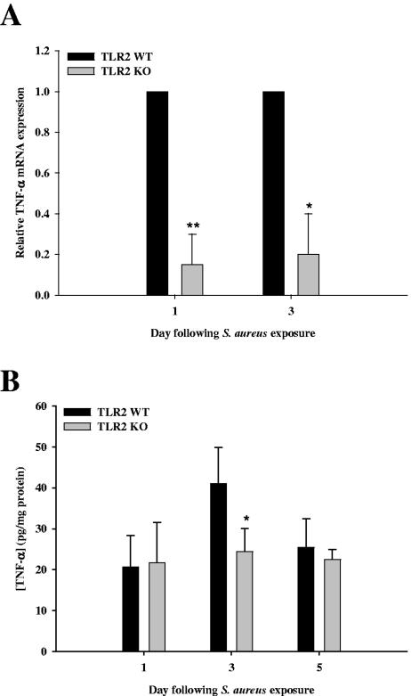
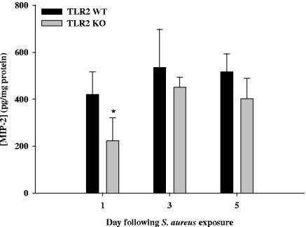
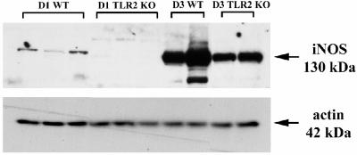
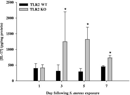
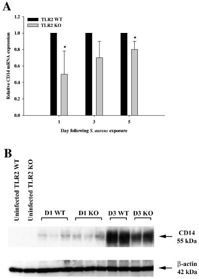
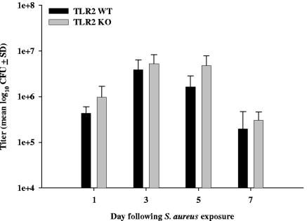
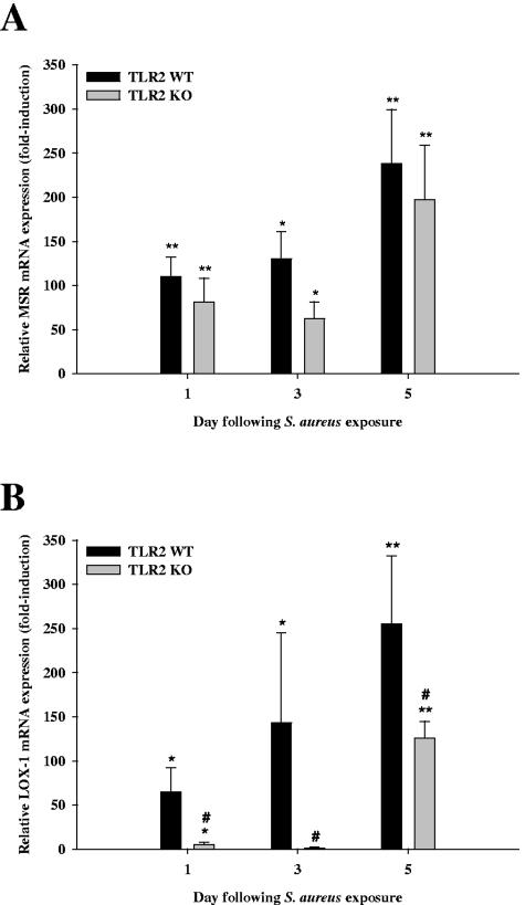
Toll-like receptor 2 (TLR2) is a pattern recognition receptor (PRR) that plays an important role in innate immune recognition of conserved structural motifs on a wide array of pathogens, including Staphylococcus aureus. To ascertain the functional significance of TLR2 in the context of central nervous system (CNS) parenchymal infection, we evaluated the pathogenesis of S. aureus-induced experimental brain abscess in TLR2 knockout (KO) and wild-type (WT) mice. The expression of several proinflammatory mediators, including inducible nitric oxide synthase, tumor necrosis factor alpha, and macrophage inflammatory protein-2, was significantly attenuated in brain abscesses of TLR2 KO mice compared to WT mice during the acute phase of infection. Conversely, interleukin-17 (IL-17), a cytokine produced by activated and memory T cells, was significantly elevated in lesions of TLR2 KO mice, suggesting an association between innate and adaptive immunity in brain abscess. Despite these differences, brain abscess severity in TLR2 KO and WT animals was similar, with comparable mortality rates, bacterial titers, and blood-brain barrier permeability, implying a role for alternative PRRs. Expression of the phagocytic PRRs macrophage scavenger receptor type AI/AII and lectin-like oxidized low-density lipoprotein receptor-1 (LOX-1) was increased in brain abscesses of both TLR2 KO and WT mice compared to uninfected animals. However, LOX-1 induction in brain abscesses of TLR2 KO mice was significantly attenuated compared to WT animals, revealing that the TLR2-dependent signal(s) influence LOX-1 expression. Collectively, these findings reveal the complex nature of gram-positive bacterial recognition in the CNS which occurs, in part, through engagement of TLR2 and highlight the importance of receptor redundancy for S. aureus detection in the CNS.
Figures
Similar articles
-
Roles of Toll-like receptor 2 (TLR2) and superantigens on adaptive immune responses during CNS staphylococcal infection.Brain Behav Immun. 2011 Jul;25(5):905-14. doi: 10.1016/j.bbi.2010.09.016. Epub 2010 Sep 22. Brain Behav Immun. 2011. PMID: 20868736 Free PMC article.
-
Toll-like receptor 2 (TLR2) is pivotal for recognition of S. aureus peptidoglycan but not intact bacteria by microglia.Glia. 2005 Mar;49(4):567-76. doi: 10.1002/glia.20144. Glia. 2005. PMID: 15593098 Free PMC article.
-
TLR2 deficiency leads to increased Th17 infiltrates in experimental brain abscesses.J Immunol. 2009 Jun 1;182(11):7119-30. doi: 10.4049/jimmunol.0802656. J Immunol. 2009. PMID: 19454709 Free PMC article.
-
Toll-like receptors in brain abscess.Curr Top Microbiol Immunol. 2009;336:41-61. doi: 10.1007/978-3-642-00549-7_3. Curr Top Microbiol Immunol. 2009. PMID: 19688327 Free PMC article. Review.
-
Forkhead Box O1 Regulates Macrophage Polarization Following Staphylococcus aureus Infection: Experimental Murine Data and Review of the Literature.Clin Rev Allergy Immunol. 2016 Dec;51(3):353-369. doi: 10.1007/s12016-016-8531-1. Clin Rev Allergy Immunol. 2016. PMID: 26924010 Review.
Cited by
-
Toll-Like Receptor 2-Mediated Autophagy Promotes Microglial Cell Death by Modulating the Microglial M1/M2 Phenotype.Inflammation. 2020 Apr;43(2):701-711. doi: 10.1007/s10753-019-01152-5. Inflammation. 2020. PMID: 31834572
-
Neuroinflammation leads to region-dependent alterations in astrocyte gap junction communication and hemichannel activity.J Neurosci. 2011 Jan 12;31(2):414-25. doi: 10.1523/JNEUROSCI.5247-10.2011. J Neurosci. 2011. PMID: 21228152 Free PMC article.
-
Blood-Brain Barrier Alterations and Edema Formation in Different Brain Mass Lesions.Front Cell Neurosci. 2022 Jul 15;16:922181. doi: 10.3389/fncel.2022.922181. eCollection 2022. Front Cell Neurosci. 2022. PMID: 35910247 Free PMC article. Review.
-
Tumor necrosis factor-alpha (TNF-alpha) regulates Toll-like receptor 2 (TLR2) expression in microglia.J Neurochem. 2007 Nov;103(4):1461-71. doi: 10.1111/j.1471-4159.2007.04838.x. J Neurochem. 2007. PMID: 17961202 Free PMC article.
-
Therapeutic Developments Targeting Toll-like Receptor-4-Mediated Neuroinflammation.ChemMedChem. 2016 Jan 19;11(2):154-65. doi: 10.1002/cmdc.201500188. Epub 2015 Jul 1. ChemMedChem. 2016. PMID: 26136385 Free PMC article. Review.
References
-
- Aggarwal, S., and A. L. Gurney. 2002. IL-17: prototype member of an emerging cytokine family. J. Leukoc. Biol. 71:1-8. - PubMed
-
- Baldwin, A. C., and T. Kielian. 2004. Persistent immune activation associated with a mouse model of Staphylococcus aureus-induced experimental brain abscess. J. Neuroimmunol. 151:24-32. - PubMed
-
- Chung, D. R., D. L. Kasper, R. J. Panzo, T. Chitnis, M. J. Grusby, M. H. Sayegh, and A. O. Tzianabos. 2003. CD4+ T cells mediate abscess formation in intra-abdominal sepsis by an IL-17-dependent mechanism. J. Immunol. 170:1958-1963. - PubMed
Publication types
MeSH terms
Substances
Grants and funding
LinkOut - more resources
Full Text Sources
Other Literature Sources
Medical
Molecular Biology Databases
Research Materials
