. 2005 Nov;115(11):3256-64.
doi: 10.1172/JCI25105. Epub 2005 Oct 20.

Lewis X component in human milk binds DC-SIGN and inhibits HIV-1 transfer to CD4+ T lymphocytes

Affiliations

Lewis X component in human milk binds DC-SIGN and inhibits HIV-1 transfer to CD4+ T lymphocytes

Marloes A Naarding et al. J Clin Invest. 2005 Nov.

Abstract

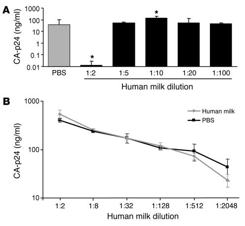
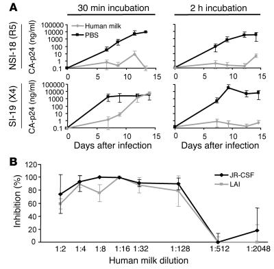
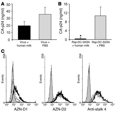
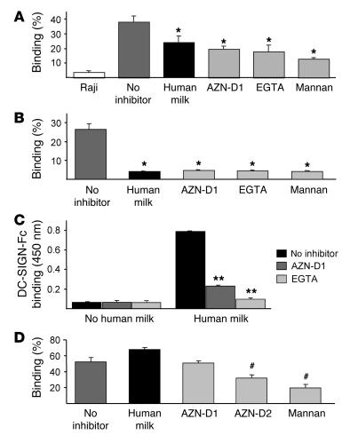
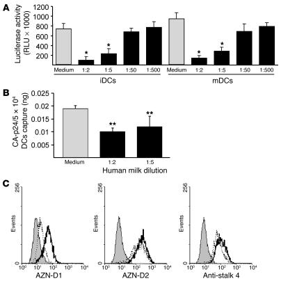
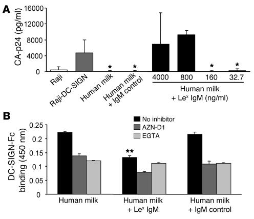
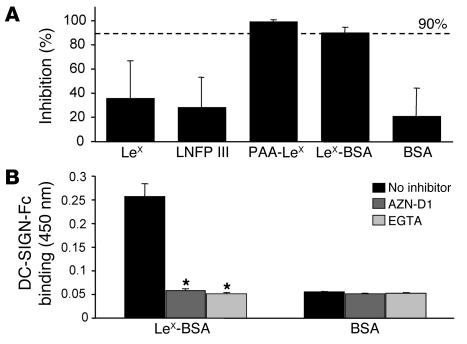
DC-specific ICAM3-grabbing non-integrin (DC-SIGN), which is expressed on DCs, can interact with a variety of pathogens such as HIV-1, hepatitis C, Ebola, cytomegalovirus, Dengue virus, Mycobacterium, Leishmania, and Candida albicans. We demonstrate that human milk can inhibit the DC-SIGN-mediated transfer of HIV-1 to CD4+ T lymphocytes as well as viral transfer by both immature and mature DCs. The inhibitory factor directly interacted with DC-SIGN and prevented the HIV-1 gp120 envelope protein from binding to the receptor. The human milk proteins lactoferrin, alpha-lactalbumin, lysozyme, beta-casein, and secretory leukocyte protease inhibitor did not bind DC-SIGN or demonstrate inhibition of viral transfer. The inhibitory effect could be fully alleviated with an Ab recognizing the Lewis X (LeX) sugar epitope, commonly found in human milk. LeX in polymeric form or conjugated to protein could mimic the inhibitory activity, whereas free LeX sugar epitopes could not. We reveal that a LeX motif present in human milk can bind to DC-SIGN and thereby prevent the capture and subsequent transfer of HIV-1 to CD4+ T lymphocytes. The presence of such a DC-SIGN-binding molecule in human milk may both influence antigenic presentation and interfere with pathogen transfer in breastfed infants.


Figures

Figure 1
Figure 1
Direct infection of CD4+ T lymphocytes in the presence of human milk. (A) PBS or several dilutions of human milk from an uninfected mother were spiked with LAI (X4) and added to CD4-enriched T lymphocytes. After a 2-hour incubation, the CD4+ T lymphocytes were washed, and fresh medium was added. (B) LAI (X4) was incubated with a 1:2 dilution of human milk or PBS for 2 hours, after which several dilutions were made and added to CD4+ T lymphocytes. For both experiments the CA-p24 concentration was determined on day 7. *P < 0.05 compared with the PBS control.
Figure 2
Figure 2
DC-SIGN–dependent transfer of HIV-1 to CD4+ T lymphocytes is inhibited in the presence of human milk. (A) A 1:2 dilution of human milk of an uninfected mother or PBS was spiked with primary isolates NSI-18 (R5) or SI-19 (X4) before addition to Raji-DC-SIGN cells. After an incubation of 30 minutes or 2 hours, the cells were washed, and activated CD4+ T lymphocytes were added. Viral replication was measured on days 7, 9, 12, and 14 after infection by determining CA-p24 values using a standard ELISA. The bars represent maximum and minimum CA-p24 values. (B) PBS or serial dilutions of human milk were spiked with JR-CSF (R5) or LAI (X4) before addition of Raji-DC-SIGN cells; after an incubation of 2 hours, the cells were washed with PBS, and stimulated CD4+ T lymphocytes were added. At day 7, CA-p24 concentrations were determined by standard ELISA. Percent inhibition was determined in reference to the CA-p24 concentration of the corresponding spiked PBS control.
Figure 3
Figure 3
The human milk compound(s) interact with the DC-SIGN receptor, which does not lead to DC-SIGN downmodulation. (A) Human milk (1:4) or PBS was preincubated with a high-titer stock of LAI before adding to Raji-DC-SIGN cells at a dilution known not to inhibit viral replication. After incubation, the cells were washed, and CD4+ T lymphocytes were added, with CA-p24 values measured on day 15 by standard ELISA (P > 0.01). (B) Human milk (1:4) or PBS were incubated with Raji-DC-SIGN, after which the cells were washed to remove unbound human milk components before addition of LAI. After incubation, the cells were washed again, and CD4+ T lymphocytes were added, with the CA-p24 values measured on day 15 by standard ELISA. *P < 0.01. (C) Raji-DC-SIGN cells were incubated with TSM or human milk (1:2) before the binding of AZN-D1, AZN-D2, and anti-stalk 4 DC-SIGN–specific Abs were determined. The filled histograms represent the isotype control; the black lines represent the Ab binding without human milk preincubation; and the dotted lines represent the Ab binding after the cells were incubated with human milk.
Figure 4
Figure 4
DC-SIGN-Fc binding ELISA and the gp120 bead adhesion assay demonstrate the interaction of the human milk compound(s) with DC-SIGN. (A and B) Raji-DC-SIGN cells or iDCs, respectively, were incubated with human milk (1:20) before addition of fluorescent gp120–coated beads. DC-SIGN–positive cells and mock Raji cells were incubated with buffer as controls. To determine the specificity of the observed binding, the cells were incubated with AZN-D1, EGTA, and mannan before addition of the gp120 beads. *P < 0.05 compared with the PBS control. (C) Human milk (1:20) was coated before addition of DC-SIGN-Fc. The specificity of the observed binding was determined by the preincubation of DC-SIGN-Fc with AZN-D1 and EGTA. **P < 0.01 compared with the noninhibitory control. (D) Raji cells expressing the L-SIGN receptor were incubated with buffer, human milk (1:20), AZN-D1, AZN-D2, or mannan before addition of the gp120 fluorescent beads. #P < 0.01 compared with the binding without an inhibitor.
Figure 5
Figure 5
Human milk inhibits the transfer of HIV-1 by iDCs and mDCs. (A) Both iDCs and mDCs from the same donor were incubated with several dilutions of human milk for 30 minutes before addition of LAI (X4). After 2 hours the cells were washed, and LuSIV cells were added; after 24 hours the LuSIV cells were washed, and the luciferase activity was determined as described in Methods. The asterisks represent statistical differences in infections (P < 0.05). (B) After an incubation of iDCs with human milk or PBS, the cells were washed and LAI was added. After an incubation of 2 hours, the cells were washed again, and captured CA-p24 levels were monitored via ELISA. **P < 0.05 compared with the corresponding control value for both experiments. (C) iDCs were incubated with TSM or human milk (1:2) before the binding of AZN-D1, AZN-D2, and anti-stalk 4 DC-SIGN–specific Abs were determined. The filled histograms represent the isotype control; the black lines represent the Ab binding without human milk preincubation; and the dotted lines represent the Ab binding after the cells were incubated with human milk.
Figure 6
Figure 6
The major milk proteins are not responsible for the inhibitory effect of human milk. (A and B) Raji-DC-SIGN cells or iDCs were incubated with the major milk proteins before addition of fluorescent gp120–coated beads; control cells were incubated with buffer. To determine the specificity of the observed binding, the cells were incubated with AZN-D1, EGTA, and mannan before addition of the gp120 beads. The asterisks represent P < 0.01 compared with noninhibitory control. (C) The major milk proteins were coated on ELISA plates, and DC-SIGN-Fc binding was measured. To determine the specificity of the observed binding, the DC-SIGN-Fc was preincubated with AZN-D1 and EGTA. *P < 0.01 compared with both the AZN-D1 and EGTA control. In all experiments the major proteins were diluted to a 1:20 dilution of their physiological concentration in human milk.
Figure 7
Figure 7
Incubation of human milk (1:20) with LeX IgM Ab relieves the inhibitory properties of human milk on DC-SIGN–mediated transfer of HIV-1 to CD4+ T lymphocytes. (A) A 1:200 dilution of human milk was incubated alone, with IgM control Ab (4,000 ng/ml), or with serial dilutions of LeX IgM Ab (4,000 to 32.7 ng/ml) before addition of Raji-DC-SIGN cells. LAI was added, and following a short incubation, the cells were washed, and activated CD4+ T lymphocytes were added, with CA-p24 values determined at day 7. *P < 0.05 compared with the Raji-DC-SIGN control. (B) Human milk (1:200) was coated and preincubated with anti-LeX IgM Ab (4,000 ng/ml) or an IgM control Ab (4,000 ng/ml) before addition of DC-SIGN-Fc to determine binding. DC-SIGN-Fc was preincubated with AZN-D1and EGTA to determine the specificity of the observed binding. **P < 0.01 compared with the human milk binding without Ab present.
Figure 8
Figure 8
Multimeric and protein-associated LeX inhibits DC-SIGN–mediated viral transfer. (A) Several LeX-containing compounds showed a difference in their ability to block DC-SIGN–dependent transfer of HIV-1 to CD4+ T lymphocytes. The LeX trisaccharide, LNFP III, PAA-LeX, LeX-BSA, and control BSA were tested in the Raji-DC-SIGN culture transfer assay at concentrations of 10 μg/ml. The inhibition is depicted as a percentage of the Raji-DC-SIGN incubated with PBS. (B) LeX-BSA and BSA as a control were coated before addition of DC-SIGN-Fc to determine the binding. DC-SIGN-Fc was preincubated with AZN-D1and EGTA to determine the specificity of the observed binding. *P < 0.01 compared with both the AZN-D1 and EGTA control.

Similar articles

Cited by

References

    1. Mitchell DA, Fadden AJ, Drickamer K. A novel mechanism of carbohydrate recognition by the C-type lectins DC-SIGN and DC-SIGNR. Subunit organization and binding to multivalent ligands. J. Biol. Chem. 2001;276:28939–28945. - PubMed
    1. Weis WI, Taylor ME, Drickamer K. The C-type lectin superfamily in the immune system. Immunol. Rev. 1998;163:19–34. - PubMed
    1. Yu Kimata MT, et al. Capture and transfer of simian immunodeficiency virus by macaque dendritic cells is enhanced by DC-SIGN. J. Virol. 2002;76:11827–11836. - PMC - PubMed
    1. Geijtenbeek TB, et al. DC-SIGN, a dendritic cell-specific HIV-1-binding protein that enhances trans-infection of T cells. Cell. 2000;100:587–597. - PubMed
    1. Pohlmann S, et al. DC-SIGN interactions with human immunodeficiency virus type 1 and 2 and simian immunodeficiency virus. J. Virol. 2001;75:4664–4672. - PMC - PubMed

Publication types

MeSH terms

Substances