. 2005 Nov;115(11):3057-71.
doi: 10.1172/JCI24792. Epub 2005 Oct 20.

Treatment of murine Th1- and Th2-mediated inflammatory bowel disease with NF-kappa B decoy oligonucleotides

Affiliations

Treatment of murine Th1- and Th2-mediated inflammatory bowel disease with NF-kappa B decoy oligonucleotides

Stefan Fichtner-Feigl et al. J Clin Invest. 2005 Nov.

Abstract

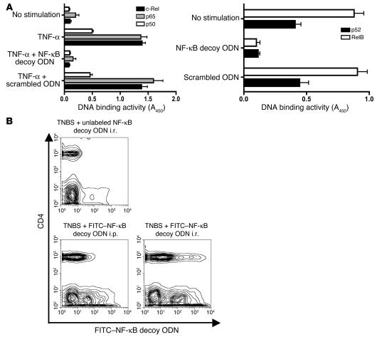
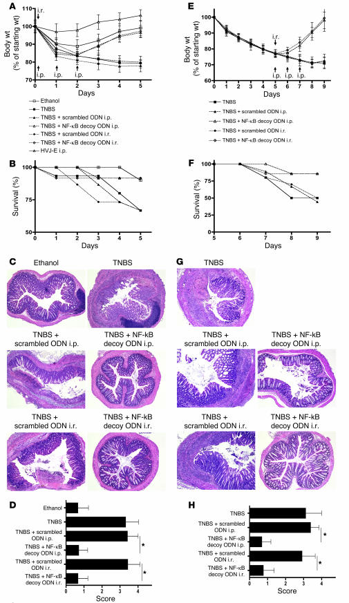
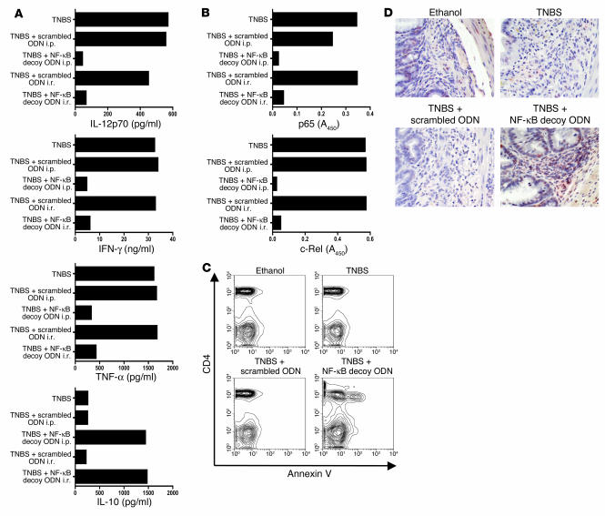
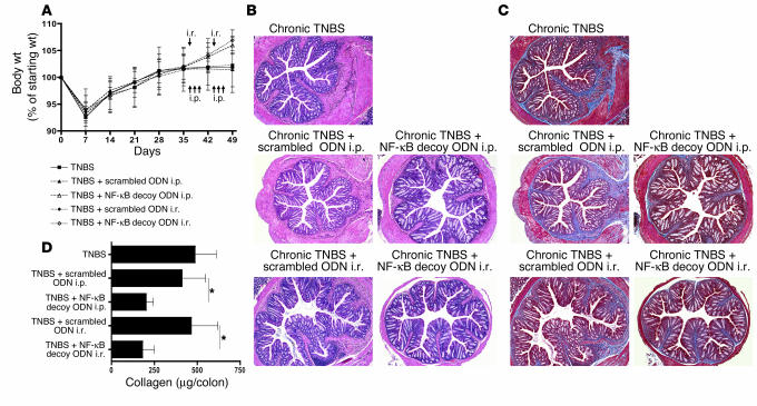
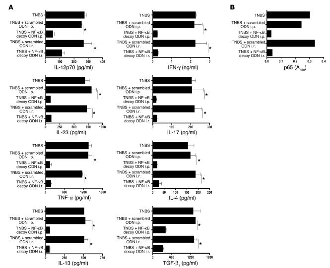
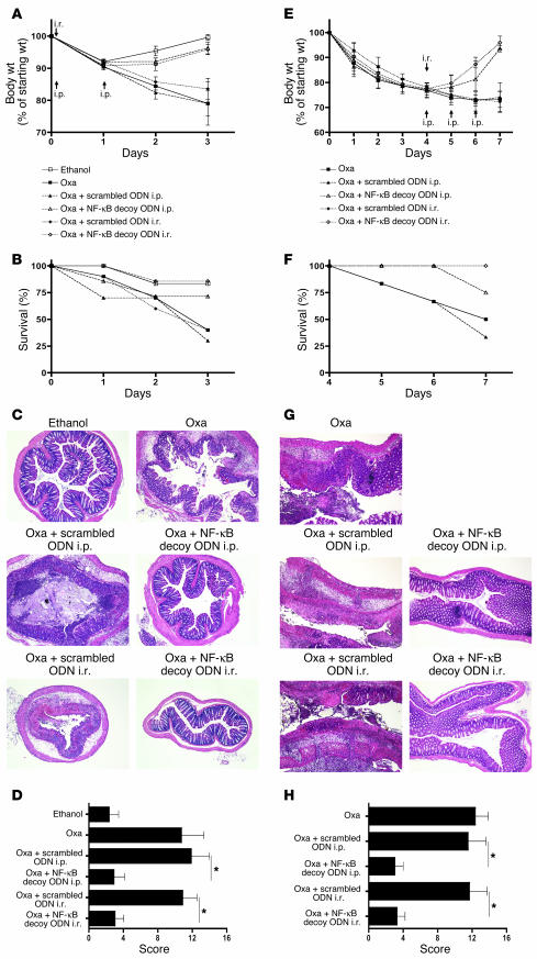
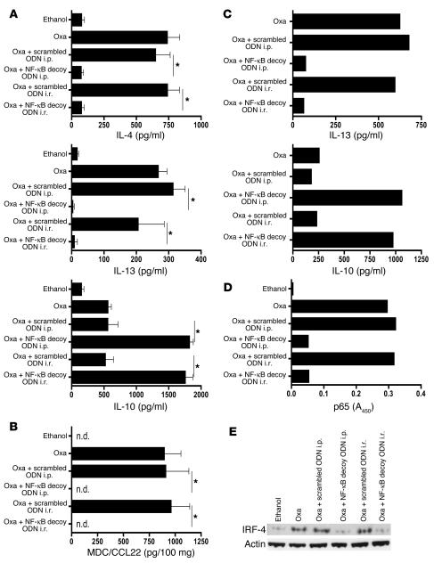
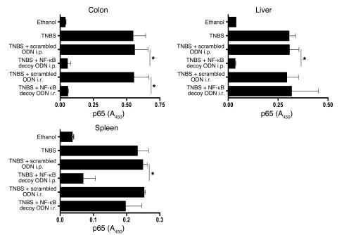
The Th1 and Th2 T cell responses that underlie inflammatory bowel diseases (IBDs) are likely to depend on NF-kappaB transcriptional activity. We explored this possibility in studies in which we determined the capacity of NF-kappaB decoy oligodeoxynucleotides (decoy ODNs) to treat various murine models of IBD. In initial studies, we showed that i.r. (intrarectal) or i.p. administration of decoy ODNs encapsulated in a viral envelope prevented and treated a model of acute trinitrobenzene sulfonic acid-induced (TNBS-induced) colitis, as assessed by clinical course and effect on Th1 cytokine production. In further studies, we showed that NF-kappaB decoy ODNs were also an effective treatment of a model of chronic TNBS-colitis, inhibiting both the production of IL-23/IL-17 and the development of fibrosis that characterizes this model. Treatment of TNBS-induced inflammation by i.r. administration of NF-kappaB decoy ODNs did not inhibit NF-kappaB in extraintestinal organs and resulted in CD4+ T cell apoptosis, suggesting that such treatment is highly focused and durable. Finally, we showed that NF-kappaB decoy ODNs also prevented and treated oxazolone-colitis and thus affect a Th2-mediated inflammatory process. In each case, decoy administration led to inflammation-clearing effects, suggesting a therapeutic potency applicable to human IBD.


Figures

Figure 1
Figure 1
Basic properties of NF-κB decoy ODNs. (A) Effect of NF-κB decoy ODNs on NF-κB DNA-binding activity. HeLa cells activated by TNF-α (20 ng/ml) or Raji cells (constitutively activated) were transfected with NF-κB decoy ODNs or scrambled ODNs encapsulated in a HVJ-E; 30 minutes after stimulation, the binding activity of p65, c-Rel, and p50 was determined in nuclear extracts of HeLa cells, whereas binding activity of Rel B and p52 was directly determined in nuclear extracts of Raji cells using the TransFactor assay. Data shown are mean values ± SD obtained from 2 independent experiments. Results are presented as absorbance at 450 nm (A450) wave length. (B) In vivo transfection of NF-κB decoy ODNs into CD4+ T cells and non-CD4+ T cells in the colonic lamina propria. Mice were administered FITC-conjugated NF-κB decoy ODNs (or unconjugated NF-κB decoy ODNs) i.r. 4 hours after TNBS administration or i.p. 4, 24, and 48 hours after TNBS administration; then, 5 days after TNBS-colitis induction, colonic lamina propria cells were isolated, stained with PE–anti-CD4, and analyzed by flow cytometry.
Figure 2
Figure 2
Treatment of TNBS-colitis by administration of NF-κB decoy ODNs. (AD) TNBS-colitis was induced by i.r. administration of TNBS in ethanol. Mice were treated with NF-κB decoy ODNs (or scrambled ODNs) via an i.r. route (at 4 hours) or via an i.p. route (at 4, 24, and 48 hours). Data shown are representative of 3 independent experiments. (A) Body weight as a percent of starting weight. Data shown are mean values ± SD and derived from at least 7 mice per group. (B) Animal survival during the first 5 days after TNBS administration. (C) H&E staining of representative colon cross-sections on day 5 after TNBS administration. Magnification, ×5. (D) Histological scores shown are mean values ± SD from at least 7 mice per group. (EH) TNBS-colitis was induced by i.r. administration of TNBS in ethanol. On day 5, mice with at least 20% body weight loss and not in recovery phase were pooled and divided into treatment groups. Mice were treated with NF-κB decoy ODNs (or scrambled ODNs) via an i.r. route (day 5) or via an i.p. route (days 5–7). Data shown are representative of 2 independent experiments. (E) Body weight as a percent of starting weight. Data shown are mean values ± SD from at least 7 mice per group. (F) Animal survival in percent until day 9 after TNBS administration. (G) H&E staining of representative colon cross-sections on day 9 after TNBS administration. Magnification, ×5. (H) Histological scores are shown as mean values ± SD from at least 7 mice per group. *P < 0.05.
Figure 3
Figure 3
Treatment of established TNBS-colitis with NF-κB decoy ODNs — effect on cytokine production, NF-κB activity, and T cell apoptosis. (AD) TNBS-colitis was induced by i.r. administration of TNBS in ethanol. On day 5, mice with at least 20% weight loss and not in a recovery phase were pooled and divided into treatment groups. Mice were treated with NF-κB decoy ODNs (or scrambled ODNs) via an i.r. route (day 5) or via an i.p. route (days 5–7). Data shown are representative of 2 independent experiments. (A) Cytokine production of colonic lamina propria cells on day 9 after TNBS administration. Cells were extracted from the lamina propria and cultured for 48 hours in the presence of stimulants (see Methods). Cytokine concentration was determined in the supernatants by ELISA. (B) DNA-binding activity of p65 and c-Rel in nuclear extracts derived from colonic lamina propria cells on day 9 after TNBS administration and measured by TransFactor assay. (C) Apoptosis of CD4+ cells in colonic lamina propria 1 day after i.r. treatment of established TNBS-colitis. Mice were treated with NF-κB decoy ODNs (or scrambled ODNs) on day 5, and colonic lamina propria cells were isolated on day 6 by flow cytometry. Apoptotic cells were determined by annexin V staining. (D) TUNEL staining of representative colon cross-sections on day 7 after TNBS administration. Magnification, ×40. Mice were treated with NF-κB decoy ODNs (or scrambled ODNs) on day 5, and colonic cross-sections were stained on day 7.
Figure 4
Figure 4
Treatment of chronic TNBS-colitis by NF-κB decoy ODNs. (AD) Chronic TNBS-colitis was induced by 7 weekly i.r. administrations of TNBS in ethanol. Mice were treated with NF-κB decoy ODNs (or scrambled ODNs) via an i.r. route (day 37 and day 44) or via an i.p. route (days 37–39 and days 44–46). (A) Body weight as a percent of starting weight. Data are shown as mean values ± SD and are representative of 2 independent experiments. (B) H&E staining of representative colon cross-sections on day 49 after TNBS administration. Magnification, ×5. (C) Masson trichrome staining of representative colon cross-sections on day 49 after TNBS administration. Magnification, ×5. (D) Collagen content of the colon. Collagen content was determined on day 49 by a Sircol assay. Data shown are mean values ± SD and are derived from at least 4 mice per group. *P < 0.05.
Figure 5
Figure 5
Treatment of chronic TNBS-colitis by NF-κB decoy ODNs — effect on cytokine production and NF-κB binding activity. (A and B) Chronic TNBS-colitis was induced by 7 weekly i.r. administrations of TNBS in ethanol. Mice were treated with NF-κB decoy ODNs (or scrambled ODNs) via an i.r. route (day 37 and day 44) or via an i.p. route (days 37–39 and days 44–46). (A) Cytokine production of colonic lamina propria cells on day 49 after the initial TNBS administration. Cells were extracted from the lamina propria and cultured for 48 hours in the presence of stimulants (see Methods). Cytokine concentrations were determined in culture supernatants by ELISA. Data shown are mean values ± SD and are representative of 2 independent experiments. (B) DNA-binding activity of p65 on day 49 after initial TNBS administration in nuclear extracts from colonic lamina propria cells, measured by TransFactor assay. *P < 0.01.
Figure 6
Figure 6
Prevention and treatment of oxazolone-colitis by administration of NF-κB decoy ODNs. (AD) Oxazolone-colitis was induced by i.r. administration of oxazolone (Oxa) in ethanol. Mice were treated with NF-κB decoy ODNs (or scrambled ODNs) via an i.r. route (4 hours) or via an i.p. route (4 hours, 24 hours). Data shown are representative of 2 independent experiments. (A) Body weight as a percent of starting weight. Data shown are mean values ± SD derived from at least 4 mice per group and are representative of 2 independent experiments. (B) Animal survival in percent until day 3 after oxazolone administration. (C) H&E staining of representative colon cross-sections on day 3 after oxazolone administration. Magnification, ×5. (D) Histological scores shown are mean values ± SD derived from at least 4 mice per group. (EH) Treatment of established oxazolone-colitis by administration of NF-κB decoy ODNs. Oxazolone-colitis was induced by i.r. administration of oxazolone in ethanol 4 days after skin presensitization with oxazolone. Mice were treated with NF-κB decoy ODNs (or scrambled ODNs) via an i.r. route (day 4) or via an i.p. route (days 4–6). (E) Body weight as a percent of starting weight. Data shown are mean values ± SD derived from at least 3 mice per group. (F) Animal survival in percent until day 7 after oxazolone administration. (G) H&E staining of representative colon longitudinal sections on day 7 after oxazolone administration. Magnification, ×5. (H) Histological scores shown are mean values ± SD derived from at least 3 mice per group. *P < 0.01.
Figure 7
Figure 7
Prevention and treatment of oxazolone-colitis with NF-κB decoy ODNs — effect on cytokine production and intracellular proteins. (A) Cytokine production in cultures of colonic LPMCs isolated on day 3 after oxazolone administration. Cytokine concentrations were determined in culture supernatant by ELISA. Data shown are mean values ± SD and are representative of 2 independent experiments. (B) Monocyte-derived chemokine/CCL22 (MDC/CCL22) production in ex vivo colon cultures isolated on day 3 after oxazolone administration; chemokine concentrations in the culture supernatant were determined by ELISA. Data shown are mean values ± SD and are representative of 2 independent experiments. (C) Cytokine production in cultures of colonic LPMCs on day 7 after oxazolone administration and day 3 after oligonucleotide administration; each culture contained cells pooled from at least 3 mice; cytokine concentrations were determined in culture supernatant by ELISA. (D) DNA-binding activity of p65 in nuclear extracts of cells derived from lamina propria on day 3 after oxazolone administration measured in nuclear extracts from colonic lamina propria cells by TransFactor assay. (E) IRF4 protein expression is reduced in colonic LPMCs. Total colonic LPMC lysates were analyzed by Western blotting on day 3 after oxazolone administration. *P < 0.01. n.d., not detectable.
Figure 8
Figure 8
Effect of NF-κB decoy ODNs administered via an i.r. route on NF-κB binding activity in extraintestinal mononuclear cells. TNBS-colitis was induced by i.r. instillation of TNBS in ethanol. Mice were treated with NF-κB decoy ODNs (or scrambled ODNs) via an i.r. route (4 hours) or via an i.p. route (4, 24, and 48 hours). DNA-binding activity of p65 on day 5 after TNBS administration was measured in nuclear extracts derived from colonic LPMCs, liver mononuclear cells, and splenocytes by TransFactor assay. Data shown are representative of 2 independent experiments involving at least 3 mice in each group. *P < 0.01.

References

    1. Podolsky DK. Inflammatory bowel disease. N. Engl. J. Med. 2002;347:417–429. - PubMed
    1. Bouma G, Strober W. The immunological and genetic basis of inflammatory bowel disease. Nat. Rev. Immunol. 2003;3:521–533. - PubMed
    1. Sartor RB. Therapeutic manipulation of the enteric microflora in inflammatory bowel diseases: antibiotics, probiotics, and prebiotics. Gastroenterology. 2004;126:1620–1633. - PubMed
    1. Maeda S, et al. Nod2 mutation in Crohn’s disease potentiates NF-κB activity and IL-1β processing. Science. 2005;307:734–738. - PubMed
    1. Heller F, Fuss IJ, Nieuwenhuis EE, Blumberg RS, Strober W. Oxazolone colitis, a Th2 colitis model resembling ulcerative colitis, is mediated by IL-13-producing NK-T cells. Immunity. 2002;17:629–638. - PubMed

Publication types

MeSH terms