. 2005 Nov;115(11):3285-90.
doi: 10.1172/JCI25247. Epub 2005 Oct 20.

P-glycoprotein deficiency at the blood-brain barrier increases amyloid-beta deposition in an Alzheimer disease mouse model

Affiliations

P-glycoprotein deficiency at the blood-brain barrier increases amyloid-beta deposition in an Alzheimer disease mouse model

John R Cirrito et al. J Clin Invest. 2005 Nov.

Abstract

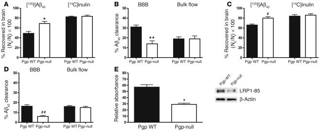
Accumulation of amyloid-beta (Abeta) within extracellular spaces of the brain is a hallmark of Alzheimer disease (AD). In sporadic, late-onset AD, there is little evidence for increased Abeta production, suggesting that decreased elimination from the brain may contribute to elevated levels of Abeta and plaque formation. Efflux transport of Abeta across the blood-brain barrier (BBB) contributes to Abeta removal from the brain. P-glycoprotein (Pgp) is highly expressed on the luminal surface of brain capillary endothelial cells and contributes to the BBB. In Pgp-null mice, we show that [I]Abeta40 and [I]Abeta42 microinjected into the CNS clear at half the rate that they do in WT mice. When amyloid precursor protein-transgenic (APP-transgenic) mice were administered a Pgp inhibitor, Abeta levels within the brain interstitial fluid significantly increased within hours of treatment. Furthermore, APP-transgenic, Pgp-null mice had increased levels of brain Abeta and enhanced Abeta deposition compared with APP-transgenic, Pgp WT mice. These data establish a direct link between Pgp and Abeta metabolism in vivo and suggest that Pgp activity at the BBB could affect risk for developing AD as well as provide a novel diagnostic and therapeutic target.


Figures

Figure 1
Figure 1
Clearance of [125I]Aβ40 and [125I]Aβ42 from the brain after intracerebral microinjection. Two- to 3-month-old WT FVB and mdr1a/b–/– mice on an FVB background were injected with 12 nmol [125I]Aβ40 or [125I]Aβ42 into the striatum. Clearance of each peptide was assessed 30 minutes after treatment. [14C]inulin, an inert reference molecule, was coinjected in each experiment to assess bulk flow elimination of ISF. (A) Brain recovery (Nb/Ni) of [125I]Aβ40 (left) and [14C]inulin (right). Significantly more [125I]Aβ40 was recovered in the brains of Pgp-null than Pgp WT mice (*P = 0.0123). Similar levels of injected [14C]inulin were recovered in both groups. (B) Calculation of [125I]Aβ40 clearance via the BBB and ISF bulk flow mechanisms (from data shown in A). Significantly less Aβ40 was cleared via the BBB in Pgp-null mice compared with Pgp WT mice (**P = 0.0033). (C) Brain recovery of [125I]Aβ42 (left) and [14C]inulin (right). More [125I]Aβ42 was cleared from the brain in Pgp WT than Pgp-null mice (#P = 0.0047). Approximately 85% of [14C]inulin was retained in both groups. (D) Similar to calculations for Aβ40 clearance, significantly less Aβ42 was cleared from the brain of Pgp-null mice (##P = 0.0004). (E) LRP1 expression was decreased by 51% in cerebral vessels of 2- to 3-month-old Pgp-null mice compared with WT FVB controls (+P = 0.002). Left: Relative absorbance representing the signal obtained from the 85-kDa LRP band on the Western blots normalized to the loading control is shown on the y axis. Right: representative lanes from an 85-kDa LRP1 peptide (LRP1-85) Western blot and protein loading control. Values are mean ± SEM; n = 4–8 per group.
Figure 2
Figure 2
Acute inhibition of Pgp increases Aβ levels in brain ISF. Using in vivo microdialysis, we assessed the concentration of ISF Aβ1–x within the hippocampus of 3-month-old APPsw mice treated with XR9576, a Pgp inhibitor. For each animal, basal concentration of ISF Aβ1–x was established over 5 hours, followed by intravenous injection of XR9576 and continued assessment of ISF Aβ for an additional 10 hours. Samples were collected at hourly intervals and assessed for Aβ1–x using a sandwich ELISA. (A) Eight hours after intravenous administration of 80 mg/kg XR9576, ISF exchangeable Aβ1–x (eAβ1–x) levels began to increase and reached 127.7% ± 3.4% of control-treated (vehicle-treated) mice (*P < 0.05; n = 5 per group). (B) Ten hours after treatment with either XR9576 or vehicle, cerebral capillaries were isolated and analyzed for levels of LRP1 expression by Western blotting (n = 5 per group). Levels of LRP1 in cerebral vessels did not change with acute inhibition of Pgp.
Figure 3
Figure 3
Biodistribution of a Pgp substrate in APPsw, Pgp-null mice. Following tail vein bolus injection of 2 μCi [99mTc]Sestamibi, brain content of [99mTc]Sestamibi in 2- to 4-month-old WT and Pgp-null mice (on an FVB background), as well as APPsw mice expressing or lacking Pgp, was determined 5 minutes after injection. More tracer was retained in all Pgp-null mice (*P = 0.0043, FVB strain; **P = 0.0058, APPsw+/– strain), demonstrating, as expected, that Pgp normally hinders the entry of [99mTc]Sestamibi into the brain. Taken together, these finding suggest that Pgp expressed on the BBB behaves similarly in WT and APPsw mice. ID, injected dose.
Figure 4
Figure 4
Increased Aβ accumulation in APPsw, Pgp-null mice. (A and B) Brain sections from APPsw mice expressing or lacking Pgp were stained with an anti-Aβ antibody. Twelve-month-old APPsw, Pgp-null (mdr1a/b–/–) mice showed greater Aβ deposition within the hippocampus and cortex than their Pgp WT littermates. Scale bar: 250 μm. (C) As quantified by unbiased stereological techniques, Pgp-null mice exhibited a greater percentage of the hippocampus covered by Aβ immunoreactivity (% Aβ load) than Pgp WT mice; **P = 0.0041. The pattern of Aβ deposition within the hippocampus was similar in both genotypes. (D) In addition, the amount of fibrillar Aβ, as assessed by the percent area of hippocampus covered by thioflavine S staining (% ThioS load), was also increased in Pgp-null animals; *P = 0.0276. (E) However, when normalized to Aβ immunoreactivity, the percentage of fibrillar Aβ to total Aβ deposits was not different between the groups; P = 0.9711. (F) Fresh hippocampal tissue was sequentially extracted with carbonate, then guanidine and Aβ ELISA was performed on the extracts. Aβ levels within each homogenate were normalized to the protein (prot.) content. Guanidine-extracted, insoluble (insol.) Aβ42 was significantly elevated in Pgp-null mice (#P = 0.0499), while there was only a trend toward elevated insoluble Aβ40 (P = 0.2452) (G). (H) Similar to the hippocampus, there was also a trend for greater Aβ immunoreactivity within the cortex of Pgp-null animals; P = 0.2788. Values are mean ± SEM; n = 11–16 per group.

References

    1. Farris W, et al. Insulin-degrading enzyme regulates the levels of insulin, amyloid beta-protein, and the beta-amyloid precursor protein intracellular domain in vivo. Proc. Natl. Acad. Sci. U. S. A. 2003;100:4162–4167. - PMC - PubMed
    1. Iwata N, et al. Presynaptic localization of neprilysin contributes to efficient clearance of amyloid-beta peptide in mouse brain. J. Neurosci. 2004;24:991–998. - PMC - PubMed
    1. Weller RO, et al. Cerebral amyloid angiopathy: amyloid beta accumulates in putative interstitial fluid drainage pathways in Alzheimer’s disease. Am. J. Pathol. 1998;153:725–733. - PMC - PubMed
    1. DeMattos RB, Bales KR, Cummins DJ, Paul SM, Holtzman DM. Brain to plasma amyloid-beta efflux: a measure of brain amyloid burden in a mouse model of Alzheimer’s disease. Science. 2002;295:2264–2267. - PubMed
    1. Shibata M, et al. Clearance of Alzheimer’s amyloid-ss(1–40) peptide from brain by LDL receptor-related protein-1 at the blood-brain barrier. J. Clin. Invest. 2000;106:1489–1499. - PMC - PubMed

Publication types

MeSH terms

Substances