. 2005 Oct 21;6(1):122.
doi: 10.1186/1465-9921-6-122.

A role for airway remodeling during respiratory syncytial virus infection

Affiliations

A role for airway remodeling during respiratory syncytial virus infection

David Becnel et al. Respir Res. .

Abstract

Background: Severe respiratory syncytial virus infection (RSV) during infancy has been shown to be a major risk factor for the development of subsequent wheeze. However, the reasons for this link remain unclear. The objective of this research was to determine the consequences of early exposure to RSV and allergen in the development of subsequent airway hyperreactivity (AHR) using a developmental time point in the mouse that parallels that of the human neonate.

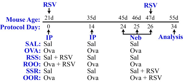
Methods: Weanling mice were sensitized and challenged with ovalbumin (Ova) and/or infected with RSV. Eight days after the last allergen challenge, various pathophysiological endpoints were examined.

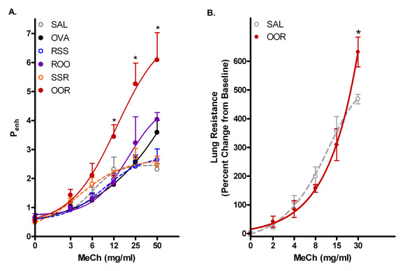
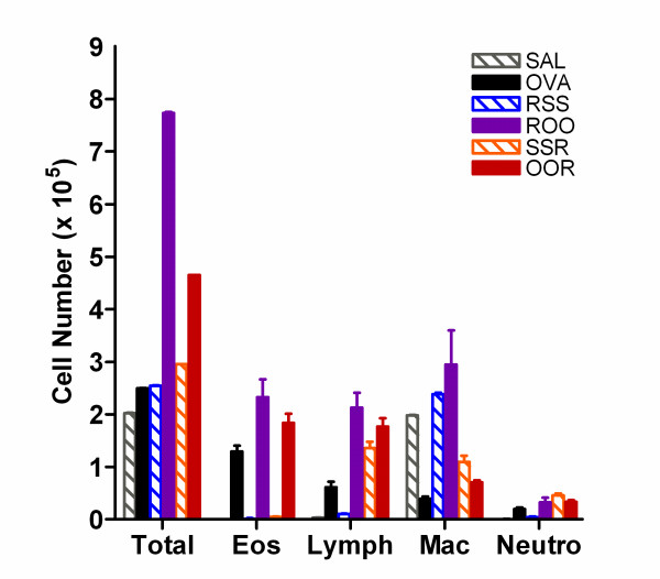
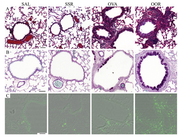
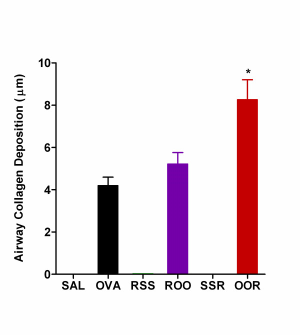
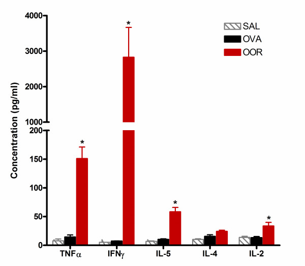
Results: AHR in response to methacholine was enhanced only in weanling mice exposed to Ova and subsequently infected with RSV. The increase in AHR appeared to be unrelated to pulmonary RSV titer. Total bronchoalveolar lavage cellularity in these mice increased approximately two-fold relative to Ova alone and was attributable to increases in eosinophil and lymphocyte numbers. Enhanced pulmonary pathologies including persistent mucus production and subepithelial fibrosis were observed. Interestingly, these data correlated with transient increases in TNF-alpha, IFN-gamma, IL-5, and IL-2.

Conclusion: The observed changes in pulmonary structure may provide an explanation for epidemiological data suggesting that early exposure to allergens and RSV have long-term physiological consequences. Furthermore, the data presented here highlight the importance of preventative strategies against RSV infection of atopic individuals during neonatal development.


Figures

Figure 1
Figure 1
Schematic of study protocol and exposure groups. Weanling mice (21 days of age) were injected i.p. with ovalbumin complexed to Imject Alum (Ova, ROO, and OOR groups) or with isotonic saline (Sal, RSS, and SSR groups) on protocol days 0 and 14. After 1 h, the ROO and RSS groups were infected with RSV (104 TCID50/g body weight). These mice were then exposed to aerosolized ovalbumin or saline for 20 minutes on protocol days 24, 25, and 26. On protocol day 26, a subset of Sal and Ova mice were infected with RSV at 104 TCID50/g body weight (SSR and OOR groups, respectively).
Figure 2
Figure 2
Enhanced airway hyperreactivity and pulmonary resistance in weanling mice exposed to RSV and OVA. A) Airway hyperreactivity (Penh) of each group is plotted as a function of increasing doses of inhaled MeCh. Data points are mean ± SEM from 8–10 mice per group. Groups as outlined in Figure 1. B) Lung resistance values were obtained by a forced oscillation technique and are plotted as a function of increasing doses of inhaled MeCh. Values presented are means ± SEM (n = 4 mice/group). *p < 0.001 for OOR vs. all other groups.
Figure 3
Figure 3
BAL fluid cellularity is altered in mice exposed to RSV and/or OVA. Differential cell counts were performed on Diff-quick stained cytospin preparations by two unbiased observers counting > 300 cells per sample. BAL cellularities are expressed as the product of the total number of cells recovered and the percentages of each cell type derived from differentials. Data are expressed as means ± SEM.
Figure 4
Figure 4
Enhanced pulmonary histopathology is observed in mice exposed to Ova and RSV. Lung sections from formalin-fixed, paraffin-embedded tissue were stained for A) cellularity, B) mucus (purple), and C) eosinophils (green) as described in the materials and methods section. The photographs are representative of the staining that occurs in the bronchioles of SAL, OVA, SSR, and OOR exposed mice. Although not shown, the RSS resembled the SSR group and the ROO group was not significantly different from the OVA mice. Scale bar = 50 μm.
Figure 5
Figure 5
Airway remodelling is evident in weanling mice exposed to Ova and RSV. Fibrosis and deposition of collagen was observed in the subepithelial, reticular layer of the airways in the OVA, ROO, and OOR mice. Morphometric analyses using data collected at 50 μm intervals over the entire basement membrane revealed that these differences were significant. (8–10 measurements at ~50 μm intervals were collected for at least 5 airways; n = 4 mice per group). ROO vs. OOR, *p < 0.05 and OVA vs. OOR, *p < 0.01.
Figure 6
Figure 6
Th1 and Th2 cytokine levels are elevated in the lungs of OOR mice. CBA analysis was used to determine the concentration of TNF-α, IFN-γ, IL-5, IL-4, and IL-2 in whole lung homogenates (n = 3 mice per group; *p < 0.01) isolated on protocol day 30. Data are expressed as means ± SEM.

References

    1. Fraenkel DJ, Bardin PG, Sanderson G, Lampe F, Johnston SL, Holgate ST. Lower airways inflammation during rhinovirus colds in normal and in asthmatic subjects. Am J Respir Crit Care Med. 1995;151(3 Pt 1):879–886. - PubMed
    1. Grunberg K, Smits HH, Timmers MC, de Klerk EP, Dolhain RJ, Dick EC, Hiemstra PS, Sterk PJ. Experimental rhinovirus 16 infection. Effects on cell differentials and soluble markers in sputum in asthmatic subjects. Am J Respir Crit Care Med. 1997;156(2 Pt 1):609–616. - PubMed
    1. Lemanske RFJ, Dick EC, Swenson CA, Vrtis RF, Busse WW. Rhinovirus upper respiratory infection increases airway hyperreactivity and late asthmatic reactions. J Clin Invest. 1989;83(1):1–10. - PMC - PubMed
    1. Glezen WP, Taber LH, Frank AL, Kasel JA. Risk of primary infection and reinfection with respiratory syncytial virus. Am J Dis Child. 1986;140(6):543–546. - PubMed
    1. McConnochie KM, Roghmann KJ. Bronchiolitis as a possible cause of wheezing in childhood: new evidence. Pediatrics. 1984;74(1):1–10. - PubMed

Publication types

MeSH terms