Review
. 2006 Feb;1762(2):232-40.
doi: 10.1016/j.bbadis.2005.06.008. Epub 2005 Sep 27.

Mitochondrial criticality: a new concept at the turning point of life or death

Affiliations
Review

Mitochondrial criticality: a new concept at the turning point of life or death

Miguel Antonio Aon et al. Biochim Biophys Acta. 2006 Feb.

Abstract

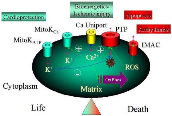
A variety of stressors can cause the collapse of mitochondrial membrane potential (DeltaPsi(m)), but the events leading up to this catastrophic cellular event are not well understood at the mechanistic level. Based on our recent studies of oscillations in mitochondrial energetics, we have coined the term "mitochondrial criticality" to describe the state in which the mitochondrial network of cardiomyocytes becomes very sensitive to small perturbations in reactive oxygen species (ROS), resulting in the scaling of local mitochondrial uncoupling and DeltaPsi(m) loss to the whole cell, and the myocardial syncytium. At the point of criticality, the dynamics of the mitochondrial network bifurcate to oscillatory behavior. These energetic changes are translated into effects on the electrical excitability of the cell, inducing dramatic changes in the morphology and the threshold for activating an action potential. Emerging evidence suggests that this mechanism, by creating spatial and temporal heterogeneity of excitability in the heart during ischemia and reperfusion, underlies the genesis of potentially lethal cardiac arrhythmias.


Figures

Fig. 1
Fig. 1
Mitochondrial inner membrane ion channels grouped according to their pro-life or pro-death effects on cardiac cells. MitoKATP: An inner membrane ATP-sensitive K+ channel activated by ischemia or K+ channel openers that is thought to play a central role in protection against ischemic injury. Multiple independent lines of evidence have characterized the properties of this channel, although the molecular structure of the pore is still unknown. MitoKCa: a toxin-sensitive (charybdotoxin, iberiotoxin) mitochondrial Ca2+-activated K+ channel has been detected in the cardiac mitochondrial inner membrane that has properties similar to KCa channels found in the plasmalemma of other cell types. KCa openers confer cardioprotection and have effects on mitochondrial function similar to those of mitoKATP channel openers. Ca Uniport: the Ca2+ uniporter is considered to be the major Ca2+ uptake pathway of the mitochondria, thus playing a role in the stimulation of NADH production and oxidative phosphorylation when workload increases. It may also contribute to mitochondrial Ca2+ overload during ischemia–reperfusion injury. PTP: the Permeability Transition Pore is responsible for the catastrophic increase in inner membrane permeability induced by Ca2+ and/or ROS. This large non-selective ion channel is capable of transporting solutes up to 1500 Da and has been linked to reperfusion injury and the induction of apoptosis. IMAC: the inner membrane anion channel is extensively discussed in the present review.
Fig. 2
Fig. 2
Mitochondrial criticality and fractal organization at the percolation threshold. (A) The mitochondrial lattice of cardiomyocytes, as imaged by two-photon laser scanning microscopy, was labeled with a membrane potential marker (top panels; TMRE labeled cell, intensity scaled from blue to white;) and a reactive oxygen species (ROS)-sensitive fluorescent probe (bottom panels; CM-H2DCFDA signal, scaled from red [reduced] to yellow [oxidized]) [28,36]. (B) The time course of ΔΨm depolarization (indicated by the arrow) and the development of the mitochondrial spanning cluster as mitochondrial ROS accumulates (lower panels of A) after a local flash (indicated by the white square). Criticality is reached when about 60% of the mitochondria have CM-DCF fluorescence levels ∼20% above baseline. This coincides with the expected theoretical percolation threshold. (C) The fractal dimension, Df, of the mitochondrial cluster, as revealed by the ROS probe (lower panels of A), was also consistent with percolation theory. Adapted from [36].
Fig. 3
Fig. 3
Mechanism of the mitochondrial oscillator. The mitochondrial and extramitochondrial processes involved in ROS-induced ROS release and the synchronization of mitochondrial activity (see text for further details). Adapted from [28].
Fig. 4
Fig. 4
Global steady-state behavior of the mitochondrial oscillator (A) and the sequence of events during an oscillatory cycle (B). (A) Bifurcation diagram of ΔΨm as a function of ROS production and scavenging. The dynamic behavior of the computational model shows an upper branch of steady states in which ΔΨm is predominantly polarized, and a lower branch, in which ΔΨm is mainly depolarized. Transitions between both branches happen at arrowheads 3 and 4. Thick lines indicate domains of stable steady-state behavior whereas thin lines denote either unstable or oscillatory states. A stable oscillatory domain, embedded within the upper branch, emerges as SOD concentration increases. Arrowheads 1 and 2 in the upper branch indicate Hopf bifurcations delimiting the oscillatory region (thin line). Adapted from [43]. (B) The time course of change in ΔΨm, IMAC flux, SOD activity, and cytosolic superoxide ([O2·−]i) during a single cycle of the mitochondrial oscillator. At a critical level of mitochondrial ROS accumulation (see Fig. 2B), the IMAC channel rapidly opens, provoking the sudden release of O2S·− from the mitochondria into the intermembrane space (see Fig. 3). The current through IMAC quickly declines due to the loss of ΔΨm. The rate of SOD increases in parallel with the burst of available O2·− and stays high until the O2S·− is consumed, at which point IMAC closes, allowing ΔΨm to repolarize, initiating the next cycle.
Fig. 5
Fig. 5
Experimental demonstration of the involvement of IMAC in ΔΨm oscillations and lack of effect by cyclosporin A. A freshly isolated cardiomyocyte loaded with the potential sensitive indicator TMRM and displaying flash-induced (arrow) mitochondrial oscillations in a continuous flow chamber, was exposed to 4-chlorodiazepam (4-Cl-DZP; 32 μM) followed by washout (W), at which time the oscillations resumed. Subsequent application of cyclosporin A (CsA; 1 μM) had no effect on the mitochondrial ΔΨm oscillations. The experimental evidence shows that, in the same cell, the antagonist of the mBzR is able to block the oscillations, and stabilizing ΔΨm in the polarized state, in a reversible manner whereas the PTP inhibitor does not. See [28] for numerous other lines of evidence excluding the involvement of PTP and Ca2+ in the response.
Fig. 6
Fig. 6
Mitochondrial oscillations drive the electrical excitability of the cardiomyocyte. (A) Action potentials (AP) (upper panel) evoked by brief current injections were recorded in current-clamp mode during whole-cell patch-clamp while simultaneously imaging ΔΨm with TMRE (lower panel). During a synchronized cell-wide depolarization-repolarization cycle, the AP shortens in synchrony with fast mitochondrial depolarization, and the cell becomes inexcitable when the mitochondria are in the depolarized state (remaining upward spikes are from the stimulus only). Recovery of ΔΨm coincided with AP restoration. (B) Correlation between the action potential duration (APD) at 90% repolarization and ΔΨm. (C) Correlation between sarcolemmal KATP current measured at 0 mV (from voltage ramps) and ΔΨm. Adapted from [28].

References

    1. Mitchell P. Coupling of phosphorylation to electron and hydrogen transfer by a chemi-osmotic type of mechanism. Nature. 1961;191:144–148. - PubMed
    1. Nicholls DG, Ferguson SJ. Bioenergetics. 3rd ed. Vol. 3. Academic Press; 2002.
    1. O'Rourke B. Pathophysiological and protective roles of mitochondrial ion channels. J. Physiol. 2000;529(Pt 1):23–36. - PMC - PubMed
    1. O'Rourke B. Evidence for mitochondrial K+ channels and their role in cardioprotection. Circ. Res. 2004;94:420–432. - PMC - PubMed
    1. Gunter TE, Pfeiffer DR. Mechanisms by which mitochondria transport calcium. Am. J. Physiol. 1990;258:C755–C786. - PubMed

Publication types

Substances

LinkOut - more resources