

Abnormalities of hippocampal surface structure in very mild dementia of the Alzheimer type
- PMID: 16243546
- PMCID: PMC2853193
- DOI: 10.1016/j.neuroimage.2005.09.017
Abnormalities of hippocampal surface structure in very mild dementia of the Alzheimer type
Abstract
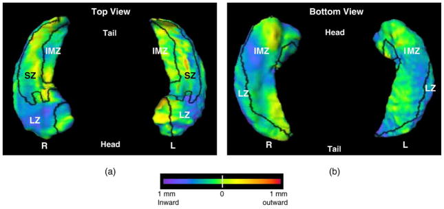
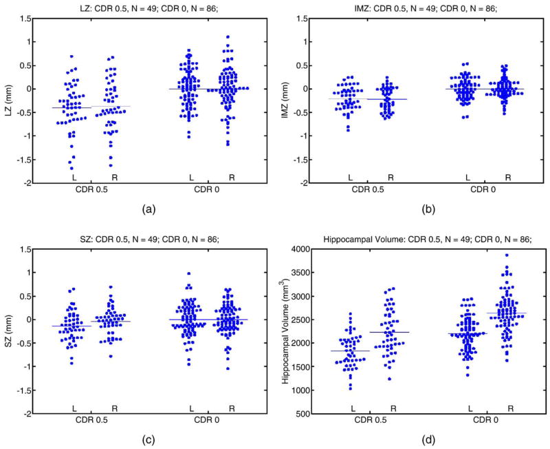
To better define the pattern of hippocampal deformity early in the course of Alzheimer's disease, we compared the pattern of hippocampal surface variation in subjects with very mild dementia of the Alzheimer type (DAT) and nondemented subjects. The surface of the hippocampus was divided a priori on a neuroanatomical template into three zones approximating the locations of underlying subfields [Csernansky, J.G., Wang, L., Swank, J., Miller, J.P., Gado, M., McKeel, D., Miller, M.I., Morris, J.C., 2005. Preclinical detection of Alzheimer's disease: hippocampal shape and volume predict dementia onset in the elderly. NeuroImage 25, 783--792]; i.e., a lateral zone (LZ) approximating the CA1 subfield, a superior zone (SZ) approximating the combined CA2, CA3, CA4 subfields and the gyrus dentatus (GD), and an inferior-medial zone (IMZ) approximating the subiculum. Large-deformation high-dimensional brain mapping (HDBM-LD) was used to generate the hippocampal surfaces of all subjects and to register the surface zones across subjects. Average variations within each zone were calculated for the subjects with very mild DAT as compared to the average of the nondemented subjects. After correcting for multiple comparisons, the very mild DAT subjects showed significant inward variation in the left and right LZ, the left and right IMZ, but not in the left and right SZ as compared to nondemented subjects. In logistic regression analyses, inward variation of the left and right LZ or IMZ by 0.1 mm relative to the average of the nondemented subjects increased the odds of the subject being a very mild DAT subject (range-1.18 to 1.57) rather than being a nondemented subject. The odds ratios for the left and right SZ were not significant. These results represent a replication of our previous findings [Csernansky, J.G., Wang, L., Joshi, S., Miller, J.P., Gado, M., Kido, D., McKeel, D., Morris, J.C., Miller, M.I., 2000. Early DAT is distinguished from aging by high-dimensional mapping of the hippocampus. Neurology 55, 1636--1643.] and suggest that inward deformities of the hippocampal surface in proximity to the CA1 subfield and subiculum can be used to distinguish subjects with very mild DAT from nondemented subjects.
Figures
Comment on
-
Preclinical detection of Alzheimer's disease: hippocampal shape and volume predict dementia onset in the elderly.Neuroimage. 2005 Apr 15;25(3):783-92. doi: 10.1016/j.neuroimage.2004.12.036. Neuroimage. 2005. PMID: 15808979
References
-
- American Psychiatric Association. Diagnostic and statistical manual of mental disorders: DSM-IV. American Psychiatric Association; Washington, DC: 1994. p. xxvi.p. 886.
-
- Analyze-AVW. Analyze-AVW. Mayo Medical Foundation; Rochester, MN: 2004.
-
- Berg L, McKeel DW, Jr, Miller JP, Storandt M, Rubin EH, Morris JC, Baty J, Coats M, Norton J, Goate AM, Price JL, Gearing M, Mirra SS, Saunders AM. Clinicopathologic studies in cognitively healthy aging and Alzheimer’s disease: relation of histologic markers to dementia severity, age, sex, and apolipoprotein E genotype. Arch Neurol. 1998;55:326–335. - PubMed
-
- Bobinski M, Wegiel J, Tarnawski M, Bobinski M, Reisberg B, de Leon MJ, Miller DC, Wisniewski HM. Relationships between regional neuronal loss and neurofibrillary changes in the hippocampal formation and duration and severity of Alzheimer disease. J Neuropathol Exp Neurol. 1997;56:414–420. - PubMed
Publication types
MeSH terms
Grants and funding
LinkOut - more resources
Full Text Sources
Medical
Research Materials
Miscellaneous
