. 2005 Nov;139(3):1207-16.
doi: 10.1104/pp.105.067520. Epub 2005 Oct 21.

The dynamic changes of tonoplasts in guard cells are important for stomatal movement in Vicia faba

Affiliations

The dynamic changes of tonoplasts in guard cells are important for stomatal movement in Vicia faba

Xin-Qi Gao et al. Plant Physiol. 2005 Nov.

Abstract

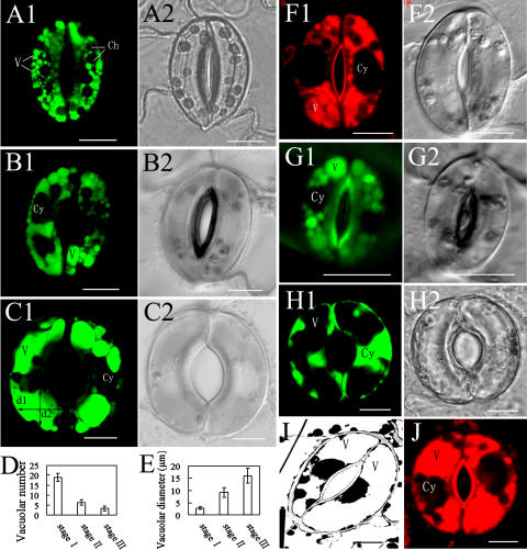
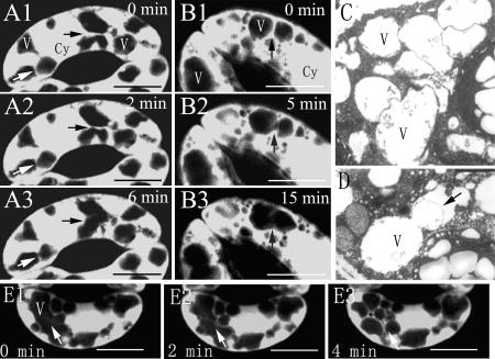
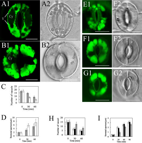
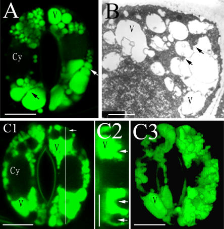
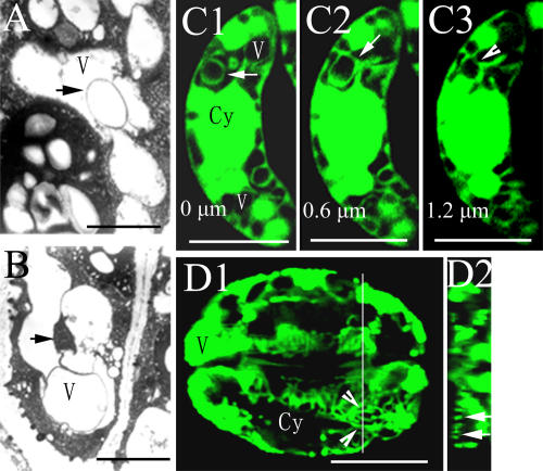
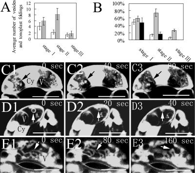
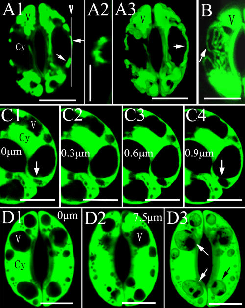
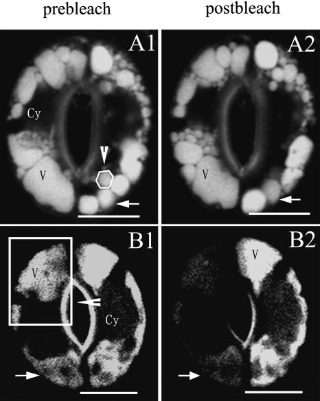
Stomatal movement is important for plants to exchange gas with environment. The regulation of stomatal movement allows optimizing photosynthesis and transpiration. Changes in vacuolar volume in guard cells are known to participate in this regulation. However, little has been known about the mechanism underlying the regulation of rapid changes in guard cell vacuolar volume. Here, we report that dynamic changes in the complex vacuolar membrane system play a role in the rapid changes of vacuolar volume in Vicia faba guard cells. The guard cells contained a great number of small vacuoles and various vacuolar membrane structures when stomata closed. The small vacuoles and complex membrane systems fused with each other or with the bigger vacuoles to generate large vacuoles during stomatal opening. Conversely, the large vacuoles split into smaller vacuoles and generated many complex membrane structures in the closing stomata. Vacuole fusion inhibitor, (2s,3s)-trans-epoxy-succinyl-l-leucylamido-3-methylbutane ethyl ester, inhibited stomatal opening significantly. Furthermore, an Arabidopsis (Arabidopsis thaliana) mutation of the SGR3 gene, which has a defect in vacuolar fusion, also led to retardation of stomatal opening. All these results suggest that the dynamic changes of the tonoplast are essential for enhancing stomatal movement.


Figures

Figure 1.
Figure 1.
Vacuoles of guard cells at different stomatal apertures. A to C, The changes of vacuolar number and size with different stomatal apertures of V. faba, stained with AO and observed by CLSM. D and E, Quantitative analysis of the average number (D) and diameter (E) of vacuoles in guard cells of V. faba at different stages of stomatal movement (at stage I, stage II, and stage III, the stomatal apertures are <5 μm, 5–8 μm, and >8 μm, respectively). Only the central optical sections of guard cells were used to calculate the average number and diameter of vacuoles. The diameter of a vacuole is the average of d1 and d2, as shown in section C1. And d1 and d2 are the largest value of vacuolar diameter paralleling with the minor axis and major axis of stomatal apparatus, respectively. More than 100 guard cells were counted at each stage. Bars represent the standard errors of three independent experiments. F, Vacuoles of guard cells of V. faba stained with LysoTracker Red DND-99, observed by CLSM. G, The result of AO staining (G1) of Arabidopsis guard cell vacuoles, observed by CLSM. H to J, Only one or two vacuoles in a guard cell of opened stomata. H, Stained with FDA and observed by CLSM; I, observed by TEM; J, stained with LysoTracker Red DND-99 and observed by CLSM. Ch, Chloroplast; Cy, cytoplasma; V, vacuole. Bars = 10 μm in A to C and F to J.
Figure 2.
Figure 2.
Fusion of small vacuoles during stomatal opening of V. faba. A and B, The fusion time-lapse images of two separated vacuoles (arrows) with stomatal opening under light, stained with FDA and observed by CLSM. Images were collected at the indicated time point. A1 to A3 were the central optical sections of stomata, and B1 to B3 were not. C, Fusing small vacuoles, observed by TEM. D, Tonoplast remnant in the lumen of two fusing vacuoles; observed by TEM. E, A big vacuole (E1, arrow) became some small vacuoles (E2 and E3, arrows) as the stomatal closing was treated with ABA, stained with FDA, and observed by CLSM. Images were collected at the indicated time point. Cy, Cytoplasma; V, vacuole. Bars = 10 μm in A, B, and E and 2 μm in C and D.
Figure 3.
Figure 3.
Fusion of small vacuoles is required for stomatal opening. A and B, Vacuoles in guard cells of V. faba present differences in number and size, with (A) or without (B) 100 μm E-64d pretreatment for 2 h, respectively, and induced 45 min under light, stained with AO, and observed by CLSM. C, The difference of stomatal opening of V. faba induced by light with 100 μm E-64d treatment for 2 h (black bar) and without 100 μm E-64d pretreatment (white bar). More than 150 stomata were counted at each stage. Error Bars represent the standard errors of three independent experiments. D, The difference of stomatal aperture of V. faba induced by cold light with 100 μm E-64d treatment (black bar) for 2 h and without 100 μm E-64d pretreatment (white bar). More than 150 stomata were counted at each stage. Error bars represent the standard errors of three independent experiments. E to G, Vacuoles in guard cells present differences in number and size of Arabidopsis mutant sgr3-1 (E), wild type (F), and complementary line sgr3-1/gSGR3 (G), after the stomata was induced 30 min under light, stained with AO, and observed by CLSM. H, The difference of vacuolar number in guard cells between Arabidopsis wild type (white bar), complementary line sgr3-1/gSGR3 (black bar), and mutant sgr3-1 (gray bar), after the stomata induced 30 min under light. More than 150 stomata were counted at each stage. Error bars represent the standard errors of three independent experiments. I, The difference of stomatal aperture between Arabidopsis wild type (white bar), complementary line sgr3-1/gSGR3 (black bar), and mutant sgr3-1 (gray bar), induced by light. More than 150 stomata were counted at each stage. Error bars represent the standard errors of three independent experiments. Cy, Cytoplasma; V, vacuole. Bars = 10 μm in A, B, and E to G.
Figure 4.
Figure 4.
Foldings of tonoplasts in guard cells of V. faba. A and B, Foldings of tonoplasts (arrows). C, Wavy surface of vacuoles. C2, The cross section of a guard cell in section C1 through the line (arrow); C3, 3D projection image of the guard cells in section C1. A and C, Stained with AO and observed by CLSM. B, Observed by TEM. Cy, Cytoplasma; V, vacuole. Bars = 10 μm in A and C and 2 μm in B.
Figure 5.
Figure 5.
Vesicle-like structures in vacuolar lumens and TVMs in guard cells of V. faba. A and B, Vesicles in vacuoles lumens (arrows), observed by TEM. C, A vesicle-like structure observed at different optical sections has a different morphology, stained with FDA and observed by CLSM. D, TVMs. D2 is the cross section of the guard cells in section D1 through the white line, showing the transection of TVMs (arrowheads) in section D1 is round (arrows), stained with AO, and observed by CLSM. Cy, Cytoplasma; V, vacuole. Bars = 2 μm in A and B and 10 μm in C and D.
Figure 6.
Figure 6.
Dynamics of tonoplast structures during stomatal movement of V. faba. A, The number of tonoplast vesicle-like structures (white bars) and foldings (gray bars) in vacuolar lumen in guard cells changed at different stages of stomatal movement (at stage I, stage II, and stage III, the stomatal apertures are <5 μm, 5–8 μm, and >8μm, respectively). Only the central optical section of a guard cell is used to count the number of foldings and vesicles. More than 180 guard cells were counted at each stage. Scale bars represent the standard errors of three independent experiments. B, The proportion of guard cells with vesicles (white bars), foldings of tonoplasts (gray bars), and TVMs (black bars) at different stages of stomatal movement (at stage I, stage II, and stage III, the stomatal apertures are <5 μm, 5–8 μm, and >8μm, respectively). More than 180 guard cells were counted at each stage. Scale bars represent the standard errors of three independent experiments. C to E, The dynamics of foldings of tonoplasts (C, arrows), vesicle-like structures (D, arrows), and TVMs (E, arrows) during stomatal opening. Time-lapse images were collected at the indicated time points. The arrows indicate moving structures. C and D, Stained with FDA and observed by CLSM. E, Stained with AO and observed by CLSM. Cy, Cytoplasma; V, vacuole. Bars=10 μm in C to E.
Figure 7.
Figure 7.
Vacuoles are interconnected in a guard cell of V. faba. A1, Two separated vacuoles (arrows). A2, A cross section image of section A1 through the white line (arrowhead), showing two separated vacuoles in section A1 are contacted. A3, The 3D projection image of the stoma in section A1, stained with AO and observed by CLSM. B, TVMs (arrow) link spherical vacuoles, stained with AO and observed by CLSM. C, Four optical sections of a stoma at different focus, showing two vacuoles are connected (arrows), stained with FDA and observed by CLSM. D, 3D projection showing interconnection of small vacuoles (arrows) in a guard cell. D1 and D2, Two optical sections of a stoma at different foci. D3, 3D projection image of the stoma in sections C1 and C2, stained with FDA and observed by CLSM. Cy, Cytoplasma; V, vacuole. Bars = 10 μm.
Figure 8.
Figure 8.
Vacuolar lumens are interconnected in a guard cell of V. faba. The photobleaching was performed with the vacuoles stained with AO bleached 50 to 100 times with strong excitation light of 488 nm under the control of LSM 510 Meta (Zeiss). Bleaching was done in the circle (in A, arrowhead) or square (in B, arrowhead). The fluorescence intensities of neighboring vacuoles (arrows) decreased after bleaching. Cy, Cytoplasma; V, vacuole. Bars = 10 μm.

References

    1. Allen GJ, Chu SP, Harrington CL, Schumacher K, Hoffmann T, Tang YY, Grill E, Schroeder JI (2001) A defined range of guard cell calcium oscillation parameters encodes stomatal movements. Nature 411: 1053–1057 - PubMed
    1. Ashford AE, Allaway WG (2002) The role of the motile tubular vacuole system in mycorrhizal fungi. Plant Soil 244: 177–187
    1. Blatt MR (2000) Cellular signaling and volume control in stomatal movements in plants. Annu Rev Cell Dev Biol 16: 221–241 - PubMed
    1. Blatt MR (2002) Toward understanding vesicle traffic and the guard cell model. New Phytol 153: 405–413 - PubMed
    1. Carter C, Pan S, Zouhar J, Avila E, Girke T, Raikhel N (2004) The vegetative vacuole proteome of Arabidopsis thaliana reveals predicted and unexpected proteins. Plant Cell 16: 3285–3303 - PMC - PubMed

Publication types

Substances

LinkOut - more resources