. 2005 Oct;1(2):e17.
doi: 10.1371/journal.ppat.0010017. Epub 2005 Oct 21.

Identification of the moving junction complex of Toxoplasma gondii: a collaboration between distinct secretory organelles

Affiliations

Identification of the moving junction complex of Toxoplasma gondii: a collaboration between distinct secretory organelles

David L Alexander et al. PLoS Pathog. 2005 Oct.

Abstract

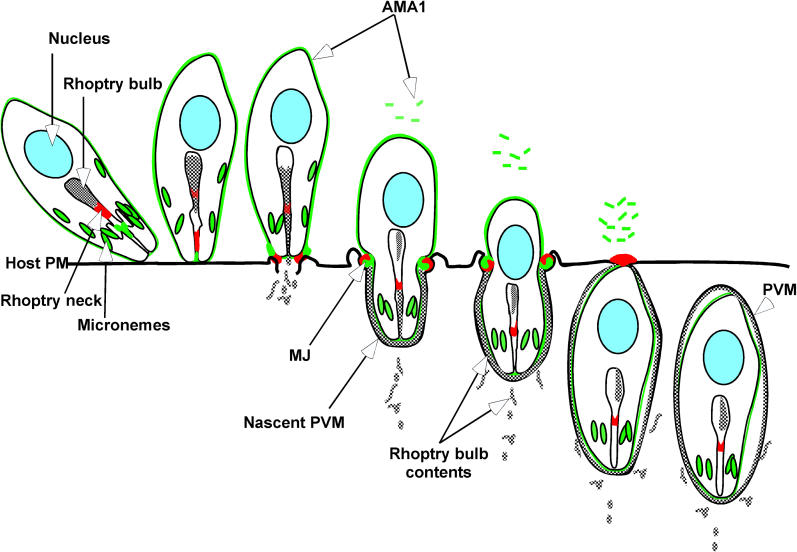
Apicomplexan parasites, including Toxoplasma gondii and Plasmodium sp., are obligate intracellular protozoa. They enter into a host cell by attaching to and then creating an invagination in the host cell plasma membrane. Contact between parasite and host plasma membranes occurs in the form of a ring-shaped moving junction that begins at the anterior end of the parasite and then migrates posteriorly. The resulting invagination of host plasma membrane creates a parasitophorous vacuole that completely envelops the now intracellular parasite. At the start of this process, apical membrane antigen 1 (AMA1) is released onto the parasite surface from specialized secretory organelles called micronemes. The T. gondii version of this protein, TgAMA1, has been shown to be essential for invasion but its exact role has not previously been determined. We identify here a trio of proteins that associate with TgAMA1, at least one of which associates with TgAMA1 at the moving junction. Surprisingly, these new proteins derive not from micronemes, but from the anterior secretory organelles known as rhoptries and specifically, for at least two, from the neck portion of these club-shaped structures. Homologues for these AMA1-associated proteins are found throughout the Apicomplexa strongly suggesting that this moving junction apparatus is a conserved feature of this important class of parasites. Differences between the contributing proteins in different species may, in part, be the result of selective pressure from the different niches occupied by these parasites.


Conflict of interest statement

Competing interests. The authors have declared that no competing interests exist.

Figures

Figure 1
Figure 1. Identification of TgAMA1-Associating Proteins
(A) Monoclonal antibodies CL22 and B3.90 were used to immunoprecipitate TgAMA1 and associating proteins from RIPA lysates of wild-type tachyzoites. The resulting material was resolved by SDS-PAGE followed by Coomassie staining to detect the proteins. Controls were immunoprecipitation with beads alone or an irrelevant mAb specific for the rhoptry bulb protein, ROP1. Each lane represents the product of 107 parasites. The polypeptides specifically associating with TgAMA1 are denoted on the right by their estimated molecular masses in kDa: p145, p130, p110, and p45. Molecular mass markers (in kDa) are indicated on the left of each panel. (B) mAb CL22 was used to immunoprecipitate TgAMA1 and associating proteins from ΔAMA/AMA1-myc parasites without (−) or with (+)Atc. (C) Immunoblot of the lanes shown in Figure 1B probed with antibody specific for TgAMA1.
Figure 2
Figure 2. Immunoprecipitation from Lysates of Parasites Subjected to Chemical Cross-Linking in Live Cultures
Tachyzoites were allowed to synchronously invade fibroblast monolayers in the absence (−) or presence (+) of the homobifunctional cross-linking agent DTSSP. Invasion and cross-linking were allowed to proceed for 15 min, then the reaction was quenched and invasion stopped. Harvested parasites were extracted in 1% SDS and heat denatured, and then processed for immunoprecipitation with antibodies to TgAMA1 (A), ROP1 (B), or RON4 (C). These immunoprecipitates were separated by reducing SDS-PAGE and analyzed by immunoblotting to determine the presence or absence of RON2, RON4, TgAMA1, or ROP1 in the co-precipitating material.
Figure 3
Figure 3. Localization of RON4 and TgAMA1 in Intracellular Tachyzoites
DIC and deconvolution processing of IIF were used to image a PV containing eight intracellular parasites that had been fixed with formaldehyde and permeabilized with triton X-100. TgAMA1 was localized with mAb CL22 and Texas-red-conjugated goat-anti-mouse antiserum. RON4 was localized via rabbit antiserum to recombinant RON4 followed by goat-anti-rabbit antiserum conjugated with fluorescein isothyocyante (FITC) (green). The images shown represent an extended focus projection through five 0.1-μm sections after iterative deconvolution. RON4 staining within the vacuole in the regions between the parasites is indicated with an arrow.
Figure 4
Figure 4. Localization of RON4 During Invasion
Tachyzoites were allowed to invade following a potassium shift as described in the text. DIC (A, E, I, M, and Q) and deconvolution IIF were then used to image formaldehyde-fixed parasites as described in Figure 3. The images presented are extended focus projections through ten 0.2-μm sections. Prior to detergent permeabilization, SAG1 was detected with the mAb DG52 followed by goat anti-mouse antiserum conjugated to FITC (green) (B, F, J, N, and R). RON4 detection was with rabbit antisera raised to recombinant RON4 and Texas red-conjugated goat-anti-rabbit antibody; this was done after permeabilization by the addition of saponin (C, G, and K) or without such permeabilization (O, S). The merged images are (D, H, L, P, and T). (A–D) represent a parasite that has just begun invasion as indicated by SAG1 staining over about 80% of the parasite. (E–H) show a parasite about halfway in. (I–L) show a parasite that appears to be fully inside (no SAG1 staining) showing small posterior cap of RON4 and apical staining consistent with rhoptry necks. (M–T) show parasites stained without prior permeabilization. (M–P) also show a parasite fully in, whereas (Q–T) show a parasite at about halfway in. The MJ is indicated by an arrow where it is clearly apparent in the DIC image.
Figure 5
Figure 5. Localization of TgAMA1 and RON4 During Invasion
Tachyzoites were allowed to invade following a potassium shift as for Figure 4. DICA, E, and I) and deconvolution IIF were then used to image formaldehyde-fixed parasites as described in Figure 3, except that the monolayers were permeabilized at the outset and a mAb specific for the cytoplasmic tail of TgAMA1 (CL22) was used in place of anti-SAG1. The images shown here are three-dimensional reconstructions from forty 0.1-μm sections. (A–D) show a wild-type parasite that is about 40% inside the host cell (the outside portion shows a bright, posterior cap of CL22 staining). (E) shows the axis of rotation for the reconstruction, and the new view of the IIF images present in (B–D) are shown in the corresponding panels (F–H). Images (I–L) show a ΔAMA1/AMA1-myc parasite with low surface AMA1 signal (no posterior cap) that is about 80% invaded into its vacuole. The MJ is indicated by an arrow.
Figure 6
Figure 6. Localization of RON4 and TgAMA1 During Egress
DIC (A) and IIF (B, C, and D) were used to image wild-type tachyzoites fixed with formaldehyde one minute after calcium-ionophore treatment to induce egress, and permeabilized with triton X-100. (B) and (C) show staining with anti-RON4 (FITC) and anti-TgAMA1 (Texas Red), respectively. (D) is the merged image. Antibody and IIF details are as for Figure 4.
Figure 7
Figure 7. RON4 localization in ΔAMA1/AMA-myc Tachyzoites Following Tetracycline Repression and Abortive Invasion
DIC (A) and deconvolution IIF (B–D) were used to image formaldehyde-fixed cultures. Images represent a projection through ten 0.2-μm sections. Intracellular ΔAMA1/AMA-myc tachyzoites were grown in Atc-containing media for 30 hr, syringe-released and then synchronized for invasion using a potassium shift as described in the methods. After allowing invasion for 20 min, the cover-slips were fixed and processed for IIF without detergent permeabilization. SAG1 staining ([B] FITC) shows an extracellular parasite in contact with the HFF monolayer. (C) shows the RON4 (Texas Red staining). (D) is a merge of (B) and (C).
Figure 8
Figure 8. Secretion and Redistribution of TgAMA1 and RON4 During Invasion
In this schematic representation, TgAMA1 (green) is secreted to the surface of parasites as they glide across the host surface. Upon reorientation, the amount of TgAMA1 on the surface is increased. A tight association with the host cell membrane is established with the secretion of RON4 (red), and rhoptry bulb constituents (grey) are detected in e-vacuoles within host cytoplasm. The MJ is established and either microneme secretion ceases or some surface proteins are able to pass through the MJ (e.g., TgAMA1, as is known for the GPI-anchored surface antigen, SAG1). RON4 migrates toward the posterior end of the parasite as a discrete ring at the constricted interface with the host cell membrane. TgAMA1 is distributed non-uniformly across the parasite surface on both sides of the MJ with a circumferential concentration at the RON4 ring. The ring containing RON4, TgAMA1, and likely RON2 and Ts4705 migrates the length of the parasite until the parasite is fully enveloped within the parasitophorous vacuole. RON4, but not TgAMA1, is then found on the host cell surface at the junction of the PV and host PM. Release of this MJ complex from the host PM allows the release of the parasite-containing PV into the host cytoplasm.

References

    1. Carruthers VB, Sibley LD. Sequential protein secretion from three distinct organelles of Toxoplasma gondii accompanies invasion of human fibroblasts. Eur J Cell Biol. 1997;73:114–123. - PubMed
    1. Mitchell GH, Thomas AW, Margos G, Dluzewski AR, Bannister LH. Apical membrane antigen 1, a major malaria vaccine candidate, mediates the close attachment of invasive merozoites to host red blood cells. Infect Immun. 2004;72:154–158. - PMC - PubMed
    1. Suss-Toby E, Zimmerberg J, Ward GE. Toxoplasma invasion: The parasitophorous vacuole is formed from host cell plasma membrane and pinches off via a fission pore. Proc Natl Acad Sci U S A. 1996;93:8413–8418. - PMC - PubMed
    1. Mordue DG, Desai N, Dustin M, Sibley LD. Invasion by Toxoplasma gondii establishes a moving junction that selectively excludes host cell plasma membrane proteins on the basis of their membrane anchoring. J Exp Med. 1999;190:1783–1792. - PMC - PubMed
    1. Aikawa M, Miller LH, Johnson J, Rabbege J. Erythrocyte entry by malarial parasites. A moving junction between erythrocyte and parasite. J Cell Biol. 1978;77:72–82. - PMC - PubMed

Publication types