Lipid interactions with bacterial channels: fluorescence studies
- PMID: 16246007
- PMCID: PMC2034676
- DOI: 10.1042/BST20050905
Lipid interactions with bacterial channels: fluorescence studies
Abstract
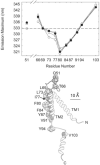
Interactions between a membrane protein and the lipid molecules that surround it in the membrane are important in determining the structure and function of the protein. These interactions can be pictured at the molecular level using fluorescence spectroscopy, making use of the ability to introduce tryptophan residues into regions of interest in bacterial membrane proteins. Fluorescence quenching methods have been developed to study lipid binding separately on the two sides of the membrane. Lipid binding to the surface of the mechanosensitive channel MscL is heterogeneous, with a hot-spot for binding anionic lipid on the cytoplasmic side, associated with a cluster of three positively charged residues. The environmental sensitivity of tryptophan fluorescence emission has been used to identify the residues at the ends of the hydrophobic core of the second transmembrane alpha-helix in MscL. The efficiency of hydrophobic matching between MscL and the surrounding lipid bilayer is high. Fluorescence quenching methods can also be used to study binding of lipids to non-annular sites such as those between monomers in the homotetrameric potassium channel KcsA.
Figures


References
-
- Lee AG. Biochim. Biophys. Acta. 2003;1612:1–40. - PubMed
-
- Lee AG. Curr. Biol. 2005;15:R421–R423. - PubMed
-
- White SH, Ladokhin AS, Jayasinghe S, Hristova K. J. Biol. Chem. 2001;276:32395–32398. - PubMed
-
- Lee AG. Biochim. Biophys. Acta. 2004;1666:62–87. - PubMed
-
- Valiyaveetil FI, Zhou Y, MacKinnon R. Biochemistry. 2002;41:10771–10777. - PubMed
Publication types
MeSH terms
Substances
Grants and funding
LinkOut - more resources
Full Text Sources
Other Literature Sources

