Case Reports
. 2005 Nov;84(11):1036-41.
doi: 10.1177/154405910508401113.

Phenotype of ENAM mutations is dosage-dependent

Affiliations
Case Reports

Phenotype of ENAM mutations is dosage-dependent

D Ozdemir et al. J Dent Res. 2005 Nov.

Abstract

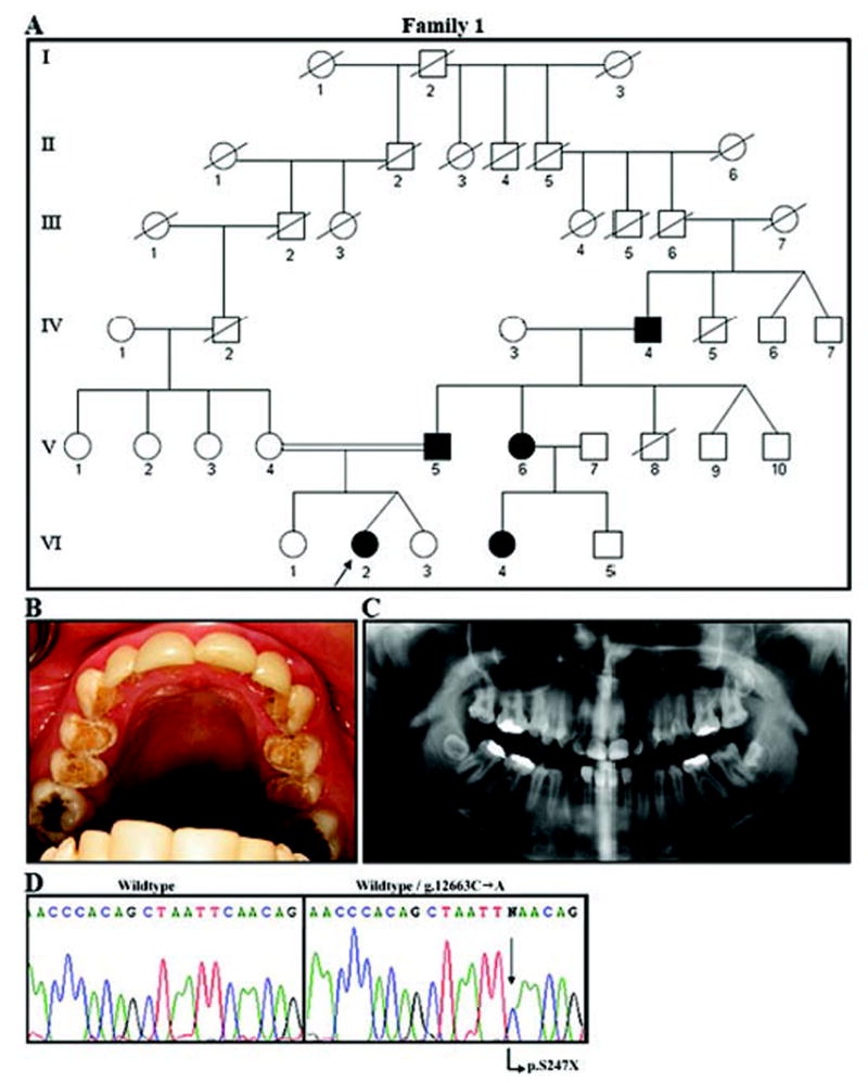
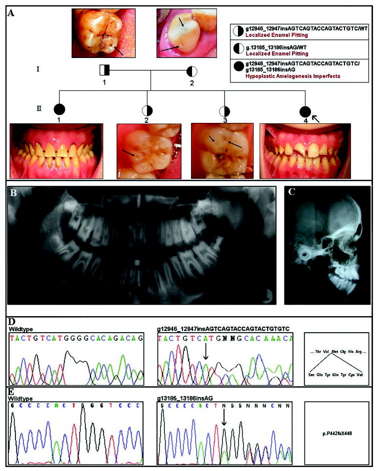
Five mutations in the ENAM gene have been found to cause hypoplastic amelogenesis imperfecta (AI), with phenotypes ranging from localized enamel pitting in carriers to severe hypoplastic AI. To determine the generality of ENAM mutations in hypoplastic AI, we sequenced the ENAM gene in ten Turkish families segregating autosomal hypoplastic AI. In two families, ENAM mutations were found. A novel nonsense mutation (g.12663C>A; p.S246X) was identified in one family segregating local hypoplastic AI as a dominant trait. Affected individuals in a second family segregating autosomal-recessive AI were compound heterozygotes for a novel insertion mutation (g.12946_12947insAGTCAGTACCAGTACTGTGTC) and a previously described insertion (g.13185_13186insAG) mutation. Heterozygous carriers of either insertion had a localized enamel-pitting phenotype. These findings substantiate that enamel phenotypes of ENAM mutations may be dose-dependent, with generalized hypoplastic AI segregating as a recessive trait and localized enamel pitting segregating as a dominant trait.


Figures

Figure 1
Figure 1
Family segregating autosomal-dominant local hypoplastic amelogenesis imperfecta. (A) Pedigree of Family 1. Blackened symbols represent AI-affected individuals. No information was available on the siblings of IV-4 or on previous generations. (B) Maxillary occlusal view of the proband (VI-2). Hypoplastic enamel is evident on the occlusal and palatal surfaces of the premolars. Laminate restorations are seen on the maxillary incisors and canines. Amalgam restorations are seen on the occlusal sites of molars. (C) The panorex radiograph (from VI-2) shows local hypoplastic enamel. Decreased contrast between enamel and dentin is evident. (D) Molecular analysis of the ENAM gene in this family. The left panel shows the wild-type sequence of a portion of exon 10. On the right is the same part of exon 10 from the proband (VI-2), showing the g.12663C>A mutation (arrow) that results in a premature stop codon (p.S246X).
Figure 2
Figure 2
Family segregating autosomal-recessive amelogenesis imperfecta. (A) Pedigree of Family 2. Right-shaded symbols indicate carriers of the 21-bp exon 10 ENAM insertion mutation. Left-shaded symbols indicate carriers of the 2-bp exon 10 ENAM insertion mutation. Individuals with either left- or right-shaded symbols have clinically evident localized enamel pitting. Representative examples of enamel pitting in these individuals are illustrated by the arrows in the photograph above or below their corresponding pedigree symbol. Completely shaded individuals are compound heterozygotes for both exon 10 ENAM mutations and have clinically apparent generalized thin hypoplastic AI. In the proband (II-4), the maxillary right incisor is fractured. All teeth are hypoplastic and yellowish, consistent with generalized hypoplastic amelogenesis imperfecta. In the affected sibling (II-1), all teeth show generalized hypoplastic yellow enamel. (B) Panorex radiograph of proband (II-4) illustrating generalized enamel hypoplasia. (C) Lateral cephalometric radiograph of individual II-4, showing anterior open bite. (D,E) Results of molecular analysis of the ENAM gene in this family. In D, the left panel shows the wild-type sequence of a portion of exon 10. The middle panel shows the same part of exon 10 from the proband (II-4), showing the g.12946_12947insAGTCAGTACCAGTACTGTGTC mutation (arrow) that results in the insertion of 7 amino acids (p.V340_M341insSQYQYCV), as shown in the right panel. In E, the left panel again shows the wild-type sequence for a portion of exon 10. The middle panel shows the same part of exon 10 from the proband (II-4), showing the previously described 2-bp insertion (indicated by the arrow; g.13185_13186insAG) that results in a frameshift and premature truncation (p.P422fsX448).
Figure 3
Figure 3
Predicted cleavage of human enamelin, based upon known porcine enamelin cleavage products. The amino acid numbers are shown above the rectangle, which represents the enamelin protein. The black rectangle represents the signal peptide (amino acids 1-39). The dotted rectangles represent cleavage products that have not been characterized at the C-terminus and are therefore shown with a question mark in the amino acid position. Enamelin is initially secreted as a 186-kDa species that undergoes rapid proteolysis to form a variety of cleavage products.

Similar articles

Cited by

References

    1. Aldred MJ, Savarirayan R, Crawford PJM. Amelogenesis imperfecta: a classification and catalogue for the 21st century. Oral Dis. 2003;9:19–23. - PubMed
    1. Backman B. Inherited enamel defects. Ciba Found Symp. 1997;205:175–182. - PubMed
    1. Bosch C, Athanasiou AE. Landmarks, variables and norms of various numerical cephalometric analyses—cephalometric morphological and growth data references. In: Athanasiou AE, editor. Orthodontic cephalometry. London: Mosby-Wolfe; 1995. pp. 241–286.
    1. Cartwright AR, Kula K, Wright JT. Craniofacial features associated with amelogenesis imperfecta. J Craniofac Genet Dev Biol. 1999;19:148–156. - PubMed
    1. Fukae M, Tanabe T, Murakami C, Dohi N, Uchida T, Shimizu M. Primary structure of porcine 89-kDa enamelin. Adv Dent Res. 1996;10:111–118. - PubMed

Publication types